Unstable Pelvic Ring Fractures

The pelvic ring consists of the sacrum and two innominate bones, which are made up of the pubis, ilium, and ischium. These bones are held together by strong ligaments to give the pelvis stability.

A pelvic ring fracture is a severe fracture with 2 breaks in the circular ring, leading to an unstable pelvis and a potentially unstable patient. Fractures that disrupt the pelvic ring predispose patients to bleeding given the large network of arterial and venous anastomoses. Patients who have an isolated pelvic fracture and are hypotensive carry a mortality of 15-40%. Most vascular injuries in the pelvis are venous (90%). While rare, arterial bleeds (10%) should be suspected when a pelvic binder is placed but the patient remains hemodynamically unstable. The retroperitoneal space can accumulate 4 liters of blood before venous tamponade occurs. Pelvic binders are useful in that they can help tamponade bleeding veins, decrease total pelvic volume, and prevent the shifting of bony fragments.

Other unstable pelvic fractures include lateral compression fractures, "open book" pelvic fractures, and vertical shear fractures. Lateral compression fractures occur when a lateral force vector (t-bone in an MVC) causes an anterior ring disruption and sacral fracture.

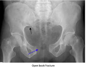
“Open book” fractures occur as a result of anteroposterior compression injury to the pelvis, commonly caused by high-speed trauma or elderly falls. There is a disruption to the pubic symphysis and the pelvis opens like a book. Diastasis of > 1 cm (blue arrow) can indicate instability. Disruption of the pubic symphysis, one of the strongest ligamentous structures in the human body, requires a lot of force and should be a red flag to look for other injuries to the head, spine, chest, or abdomen.

Vertical shear pelvic fractures are seen when one-half of the pelvis shifts upward as a result of a fracture of ipsilateral anterior and posterior pelvic ring fractures. They typically occur as a result of high-energy force applied in the axial direction (aka from the gas pedal to the femur and up to the pelvis). Patients may have an unstable pelvis and leg length discrepancy.

For all unstable fractures, you should appropriately resuscitate and stabilize the patient. Give blood as needed but avoid transfusing through lower limb access because it may drain into the retroperitoneal space. If there is a pelvic ring fracture, consider binding the pelvis. Your binder should lay over the greater trochanters and have enough force to close the pelvic ring (video:https://www.youtube.com/watch?v=tWLBZKeWEkg).


Acute Compartment Syndrome

Acute compartment syndrome is when the pressure in a muscle compartment increases, compromising circulation and function. This occurs because the compartment is surrounded by a fascial membrane that restricts further expansion. It typically occurs after trauma, crush injury, or burns. Signs include severe pain (earliest sign), pallor, paresthesia, paresis, and pulse deficit. To measure compartments, you take your measurement device and insert it into the compartment of interest. 

How to set up your measurement device: 

  • Your materials include a sterile 3cc saline syringe, chamber, and needle. The needle has a side port (hole) for measuring pressure.

  • Connect the syringe, chamber, and needle

  • Flush the chamber and needle with saline to get rid of the air; do this by holding the entire device at a 45-degree angle.

  • Load into the monitoring unit and press zero, you should see 00

  • Insert the needle into the compartment of choice and hold it for reading

There are two ways to assess for compartment syndrome. You can use the absolute or delta pressure (normal: 0-8). Suspect compartment syndrome if:

  • the absolute pressure is > 30 mmHg

                                   OR

  • The delta pressure is < 30 mmHg

    • Delta pressure = diastolic pressure - compartment pressure. This means that the pressure in your compartment is so high that it is close to your diastolic blood pressure

Tip: remember the number 30 

These patients require a fasciotomy so call ortho ASAP. Meanwhile, you should level their affected limb and support BP if hypotensive to help maintain perfusion. 

Thanks for reading!

-Ariella

References: 

https://www.emrap.org/episode/trauma1/compartment

https://www.emrap.org/episode/measuring/measuring


POTD: Listen, Frank, let's talk about Lisfranc Injuries

Background

Lisfranc ligament attaches 2nd metatarsal to medial cuneiform

2nd metatarsal is held in mortice created by the three cuneiform bones

Injury to 2nd metatarsal often results in dislocation of the other MTs

Dorsalis pedis may be injured in severe dislocation

Lisfranc Injury = any fracture or dislocation of the tarsal-metatarsal joint

Mechanism of Injury

MVAs, falls from height, and athletic injuries

Indirect rotational forces and axial load through hyper-plantarflexed forefoot

hyperflexion/compression/abduction moment exerted on forefoot and transmitted to the TMT articulation

metatarsals displaced in dorsal/lateral direction

Clinical Features

Inability to bear weight (especially on tiptoe)

Tenderness over tarsometatarsal region

Pain with pronation and passive abduction of the midfoot

Ecchymosis of plantar section of midfoot is highly suggestive

Imaging

Obtain radiographs, which include AP, lateral, oblique, and weight bearing views.

AP: Medial margin of 2nd metatarsal base does not align with medial margin of 2nd cuneiform. Bony displacement 1mm or greater between bases of first and second metatarsals is considered unstable.

Oblique: Medial margin of 3rd metatarsal does not align with medial margin of 3rd cuneiform.

Lateral: 2nd metatarsal is higher than middle cuneiform (step-off).

If suspicion is high based on history and physical, you may want to consider obtaining further imaging in conjunction with your ortho consultants.

Treatment and Dispo

Sprains and non-displaced fractures:

Non-weightbearing splint with ortho follow up (most managed with cast x 6 weeks)

Posterior Ankle Splint

Displaced fractures:

Emergent ortho consult

When diagnosed appropriately, patients who undergo open reduction and internal fixation of fractures have superior outcomes to those with purely ligamentous injury

20% are missed on first presentation to ED, so keep this in mind the next time you see a patient with the chief complaint of foot pain!

References:

https://www.orthobullets.com/foot-and-ankle/7030/lisfranc-injury

Sherief, T et al. Lisfranc injury: How frequently does it get missed? And how can we improve? Injury: International Journal of the Care of the Injured 2007: 34; 856-860. PMID: 17214988

 ·