STEMI Equivalents

STEMI Equivalents

Hyperacute T Waves

  • in ≥ 2 contiguous leads

  • broad and asymmetric

  • concerning when upright T-wave in V1 > V6

  • also need to rule out hyperK

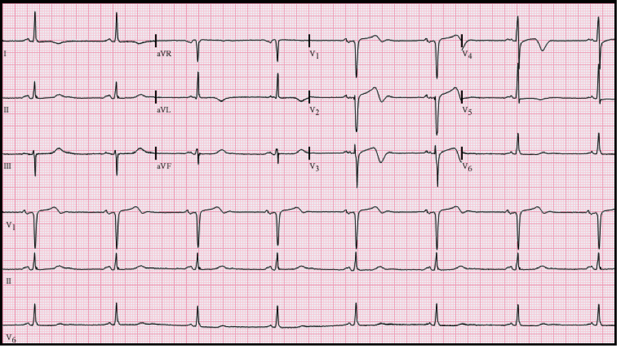
Wellen's Waves

  • Commonly in V/2/V3

  • Type A: biphasic T waves

  • Type B: more common; deep, symmetric T waves inversions

ekg2.png

AVR

  • ST Elevation >1mm in AVR and/or V1 with diffuse depressions

  • LMCA occlusion, LAD occlusion, or triple vessel disease

    • Nonspecific, can also see this EKG with PE, aortic dissection, etc.

ekg4.png


De Winter’s T Waves

  • ST depression with peaked T Waves

    • > 1 mm of upsloping ST depression and tall symmetric T-waves commonly in the precordial leads

  • Proximal LAD occlusion

ekg6.png

Also consider:


Posterior MI

  • ST depressions in V1-V3

  • Get posterior EKG with leads V7V8V9

    • STE ≥ 0.5 mm (≥ 1 mm in men < 40 years) in V7, V8, or V9

  • Left circumflex or RCA occlusion

Right Ventricular MI

  • should consider in your inferior STEMI pts

  • ST elevation in V1 (+/- V2 ST elevation OR depression)

  • ST elevation in lead III > lead II

  • Get a right sided EKG with leads V3V4V5

    • Can have elevations in these

  • RCA occlusion

 · 
Share

Ventriculoperitoneal  Shunt (Complications)

Background

  • Placed in the management of hydrocephalus

    • Hydrocephalus can be secondary to many disease processes, some included below:

      • Congenital

      • Spina bifida

      • Tumors

      • Post-meningitic

      • Dandy walker syndrome

      • Arachnoid cysts

      • Idiopathic Intracranial HTN

  • Location of the shunt is based on the location of blockage causing the hydrocephalus

    • Ventricular catheter can be placed in any brain ventricle (lateral, third, fourth)

      1. Valve portion then connects to distal end of the catheter/tubing, which can terminate in tissue that has epithelial cells capable of absorbing incoming CSF

        1. Most commonly in the abdominal peritoneal space, but can also be placed in the heart (right atrium, VA shunt), pleural cavity, etc. (see below images)

  • Most common neurosurgical procedure to cause complications

  • shunt failure occurs in 14% of children in the first month, 50% in first year

Symptoms of Complications

  • Adults: nausea/vomiting, lethargy, AMS, ataxia, CN palsies, paralysis of upward gaze (“sunset eyes”), seizures

  • Children: nausea/vomiting, irritability, lethargy, change in behavior, seizures, bradycardia, apnea, bulging fontanelle, prominent scalp veins,


Under-shunting

  • Obstruction of shunt flow

  • Develop high ICP and then aforementioned symptoms

  • Can be caused by extra-luminal obstruction or intraluminal obstruction

  • Extra-luminal obstruction

    • disconnection, kinking or fracture of the shunt system

  • Intra-luminal obstruction

    • Blockage caused by blood or CNS/inflammatory cells secondary to infection or tumor


Over-shunting

  • Over-drainage of CSF

  • Develop intracranial hypotension aka low ICP

    • Siphoning effect of CSF fluid upon standing

    • Develop headache that’s relieved in recumbent position

  • Can lead to slit ventricles

    • Complete collapse of the ventricles

    • Most patients are asymptomatic

    • Few will develop Slit Ventricle Syndrome

      1. Pathophys not fully understood

  • Can cause subdural hematoma

    • Over-shuntingàbrain collapseà tearing of bridging veins


CSF Shunt Infection

  • Usually within 6 months of placement

  • Can have fever, but not mandatory

  • External Infection = subcutaneous tract around the shunt

    • Swelling, erythema, tenderness along area of shunt tubing

  • Internal Infection = shunt and CSF contained within the shunt

    • Symptoms above

  • Staph epidermidis (50%) > Staph aureus (20%) > gram-negative rods (15%) > Propionibacterium acnes

  • Require shunt tap, usually by neurosurgery . not LP!

  • AB = cephalasporin + vanc


Work up

  • Labs are not very helpful

    • Can get cbc, sed rate, blood cultures

  • CSF

    • Protein can be high

    • Glucose can be low

    • Cultures negative 40% of time

  • Shunt series

    • XRs along course of VP shunt

    • Useful to visualize fractures/disconnection/migration of tubing (see below images)

    • Compare to old series

vp5.png

    • Just because shunt series may show a disconnection doesn’t mean theres actually a malfunction.

      1. Shunt may still be draining csf through another tract

  • CT head (non-con)

    • Should be paired with shunt series to further asses for malfunction

      1. Should not obtain shunt series/CT alone, should always be paired with each other

  • MRI

    • Interestingly, shunt hardware difficult to evaluate on mri

  • VP shunt tap

    • Indications in chart below

    • Almost always done by neurosurgery

  • Medications

    • Symptomatic therapy (Zofran, pain control etc)

    • if suspect infxn, AB as stated above

    • Consult with neurosurgery about starting steroids/acetazolamide to reduce ICP

  • Dispo

    • If presentation/imaging concerning then admit for further neurosurgery follow up

https://wikem.org/wiki/Ventriculoperitoneal_shunt_problems

http://www.emdocs.net/complications-csf-shunts-ed-presentations-evaluation-management/

https://www.ncbi.nlm.nih.gov/books/NBK459351/


 · 
Share

Supracondylar Fractures

sc9.png

Pediatric Supracondylar Fractures

Background

  • Defined by fracture of distal aspect of humerus above the epicondyles

  • Mechanism

    • Direct: blow to the elbow, fall onto flexed elbow

    • Indirect (more common): FOOSH, fall onto hyperextended UE

      1. 95% of these fractures are due to extension injury

  • Most common age: 5-8 year olds

    • Also more likely to be dislocated in this age group

  • Males>Females

Exam

  • Complain of pain/swelling/decreased ROM of elbow

  • “S shaped deformity”

    • when fracture is entirely displaced (distal humerus)

  • Need to perform neurovascular exam!

    • Median nerve: A-OK sign

      1. Mostly commonly affected

    • Radial nerve: thumbs up sign

    • Ulnar nerve: abduct/adduct fingers (try to remove paper they are holding in between adducted fingers)

  • Check for cap refill!

  • Evaluate brachial artery

    • Compromise of the artery can lead to permanent volkmans contracture, which is flexion at the wrist

Gartland Classification

  • Based on the integrity of the cortex and extent of displacement

  • Type 1: minimal to no displacement ; limited XR findings, look for occult signs of fx on xray (ie: fat pad)

  • Type 2: posterior hinge aka displaced anterior wall but intact posterior wall; anterior humeral line is anterior to capetellum

  • Type 3: complete displacement with no cortices in tact, neither anterior nor posterior wall in tact

  • Type 4: periosteal disruption with instability in extension AND flexion

sc5.png

Imaging

  • Need AP and lateral films

  • Lines

    • Abnormality can indicate occult fracture

    • Radiocapitellar line (yellow): Line through central radius and central capitellum (middle third). Should be evaluated in both views

    • Anterior humeral line (blue): Line in front of the humerus and passes the anterior 1/3 of the capitellum.

  • Fat pads

    • Anterior: can be normal; elevation is abnormal

    • Posterior: always pathologic

  • These abnormalities without obvious sign of fracture along bones indicative of type 1 SC fx

Dispo:

  • Type 1: long arm posterior splint, ortho follow up

  • Type 2/3: OR with ortho for reduction (closed vs. open) and pinning


 · 
Share