HiNTS exam

What

Series of three quick, bedside, physical exam maneuvers that can potentially rule out a central cause of vertigo


Hi for head impulse testing, or head thrust testing.
N for nystagmus to remind you to look for direction-changing or vertical nystagmus
TS for test of skew.


HINTS1.png


Why

  • Nearly two-thirds of patients with stroke lack focal neurologic signs that would be readily apparent to a nonneurologist

  • Presence of all three “reassuring” exam findings suggests it can be ruled out with a 100% sensitivity for ischemic stroke in AVS while an initial MRI with diffusion-weighted imaging (DWI) had a 88% sensitivity


Who

Maneuvers used to help distinguish between central and peripheral vertigo in patients experiencing an acute vestibular syndrome (AVS) which is best defined as: rapid-onset vertigo, nausea and/or vomiting, gait unsteadiness, head motion intolerance, and nystagmus.


When

The patient must be experiencing continuous vertigo for the results to be reliably interpreted.

How

Head Impulse Test

HINTS2.png


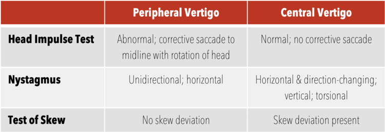
  • Ask the patient to relax his/her head and maintain his/her gaze on your nose. Gently move the patient’s head to one side, then rapidly move it back to the neutral position. The patient may have a small corrective saccade. The head impulse test is positive (consistent with peripheral vertigo) if there is a significant lag with corrective saccades. If you can see the correction, it is abnormal. Compare this to the contralateral side; a difference in the speed of correction should be noted.

  • In acute vestibular syndrome, an abnormal result of a head impulse test usually indicates a peripheral vestibular lesion, whereas a normal response virtually confirms a stroke.

  • Abnormal exam rules in peripheral vertigo and thus rules out central vertigo if only unilateral

  • Video- https://www.youtube.com/watch?v=XpghlvnrREI&feature=youtu.be&t=665


Nystamus

  • Note if it is present in primary gaze (i.e. looking straight ahead) and or in lateral gaze. Unidirectional, horizontal nystagmus is reassuring for peripheral vertigo where as purely bidirectional, vertical or torsional can be consistent with central cause

  • The most common peripheral nystagmus, BPPV, in the posterior semicircular canal consists of a unidirectional horizontal nystagmus with a torsional component.


Test of Skew

  • Have the patient maintain his/her gaze on your nose. Alternate covering each of the patient’s eyes

  • Positive result will be the deviation of one eye while it is being covered, followed by correction after uncovering it.

https://www.youtube.com/watch?v=WAPaIMMsV_A


Summary

  • If the HiNTs exam is entirely consistent with peripheral vertigo (positive head impulse test, unidirectional and horizontal nystagmus, negative test of skew), then, according to the derivation paper, it is 100% sensitive and 96% specific for a peripheral cause of vertigo.

  • Use of HiNTs exam in the ED is currently controversial as the primary study was performed by neurologists in a partially differentiated patient population

  • likely has higher utility in the patient population in whom the clinician suspects a peripheral cause of their vertigo to rule out central cause and limit needless imaging


Limitations

  • Do not perform on any patient that has head trauma, neck trauma, an unstable spine, or neck pain concerning for arterial dissection.

  • Do not perform in patients with known severe carotid stenosis as it may embolize unstable plaque

  • Challenging to differentiate between catch up saccade and nystagmus

  • Patients with acute active AVS likely to not tolerate the testing

  • Patient must be awake and cooperative.

  • Essentially an awake “doll’s eye” that requires conscious fixation on an object. Cannot perform on mentally impaired or sedated patients

  • Not yet been validated by a large external group, let alone a large external group of emergency medicine providers.

  • In the study, exam performed by ophtho neurologists


References


NUEMBlog

Tamingthesru

Nelson, James A., and Erik Viirre. "The clinical differentiation of cerebellar infarction from common vertigo syndromes." Western Journal of Emergency Medicine 10.4 (2009): 273.

Kattah, Jorge C., et al. "HINTS to diagnose stroke in the acute vestibular syndrome: three-step bedside oculomotor examination more sensitive than early MRI diffusion-weighted imaging." Stroke 40.11 (2009): 3504-3510.

Tarnutzer, Alexander A., et al. "Does my dizzy patient have a stroke? A systematic review of bedside diagnosis in acute vestibular syndrome." CmAJ 183.9 (2011): E571-E592.


 · 

Metformin Associated Lactic Acidosis 


Metformin Associated Lactic Acidosis 

"The dose makes the poison"

Metformin a common medication used to treat DM II can cause metformin-associated lactic acidosis and Metformin toxicity with a mortality rate between 45-48%. Neither arterial lactate levels nor plasma metformin concentrations predicted mortality.

met cycle.jpg


  • · The main effect of metformin is inhibition of the mitochondrial transport chain complex-I, which essentially poisons the mitochondria.

  • · If the mitochondrial transport chain stops working: NADH builds up, Krebs cycle eventually gets backed up, Pyruvate gets converted into lactate which builds up.


Best described a spectrum depending on cause of lactic acidosis


Metformin-induced lactic acidosis (MILA)


  • · High levels of metformin are the primary cause of illness.


Acute metformin overdose


    • · Acute poisoning may lead to MILA in the absence of renal dysfunction.

    • · Precise amount of metformin required to do this is unclear, but seems to be high (e.g. >20 grams).

    • · Patients with acute ingestion look fine initially, but deteriorate subsequently (“toxin bomb”).

Subacute accumulation of metformin due to renal failure


    • · Metformin is renally cleared

    • · Progressive renal failure (with GFR << 30 ml/min) eventually leads to metformin accumulation and toxicity.

    • · These patients may present with marked lactic acidosis, yet have fairly preserved hemodynamics and look OK.


Metformin-associated lactic acidosis (MALA)

Patient on metformin develops an acute life-threatening illness (e.g. septic shock, cardiogenic shock). Metformin amplifies the degree of lactic acidosis, but it's not the sole cause of the illness. Risk factors include renal insufficiency, higher doses of metformin, and alcoholism.


Metformin-unrelated lactic acidosis (MULA)


  • · Metformin levels are low; metformin is an innocent bystander.

  • · Clinically it will be impossible to differentiate this from MALA. Differentiation of MULA from MALA requires measurement of metformin levels, which isn't available at most hospitals.


Signs & symptoms


  • · Vitals: The following abnormalities may be seen: Hypothermia, Hypotension progressing to vasopressor-refractory shock can occur.

  • · GI symptoms often predominate: Nausea, vomiting, diarrhea, epigastric pain.

  • · Delirium, decreased consciousness


Management


  • · Fingerstick glucose (hypoglycemia may occur)

  • · VBG with lactate

  • · Complete set of chemistries (including Ca/Mg/Phos), Coags

  • · Beta-hydroxybutyrate level (frequently elevated)

  • · Liver function tests

  • · Blood cultures, urinalysis, chest X-ray, procalcitonin.

  • · Administer Broad-spectrum empiric antibiotics

  • · Additional toxicologic evaluation (e.g. acetaminophen, salicylate levels, toxic alcohols, carboxyhemoglobin).

  • · Obtaining a serum metformin concentration is unhelpful in most cases because few hospitals perform the test and thus timely results are rarely available, and because the serum concentration often does not correlate with the severity of the poisoning or patient outcome

  • · Obtain early consultation with a medical toxicologist and a nephrologist


Metformin-induced lactic acidosis vs. DKA


  • · Compared to isolated DKA, patients with metformin-induced lactic acidosis have greater degree of hyperlactatemia, with less extensive ketoacidosis.

  • · Difficult to sort this out in some situations- treat both conditions (the treatment for DKA may actually improve MILA/MALA).

  • · Activated charcoal may be considered for patients who present very shortly following acute ingestion, without contraindications (e.g. normal mental status without risk of aspiration)

  • · Evaluate for alternative causes of illness, especially septic shock.

  • · Insulin therapy may be beneficial for metformin poisoning (aside from any question of DKA) by reducing generation of lactate and ketosis thereby improving acidosis


Bicarbonate?


  • · Undesirable for a few reasons: Might increase cellular permeability to metformin, Bicarbonate has never been shown to be a useful therapy for lactic acidosis, Raising the pH with bicarbonate may actually stimulate glycolysis and thereby increase lactate generation

  • · Normal saline is an acidotic fluid that will exacerbate the acidosis.

  • · Lactated Ringers and Plasmalyte not good choice, as these patients cannot metabolize lactate or acetate respectively


Other options?


  • · D5W with 1/2 normal saline, plus one ampule (50 mEq) of bicarbonate added per liter.

  • · Simultaneous infusions of normal saline and isotonic bicarbonate

  • · High-flow nasal cannula may be used to improve ventilatory efficiency and reduce the work of breathing


Indications for dialysis


  • · Main indications

    • · Lactate >15-20 mM

    • · pH <7.0-7.1

    • · Failure to improve despite standard supportive measures

  • · Comorbid conditions which may lower the threshold for dialysis

  • · If the patient is hemodynamically unstable, CVVH or CVVHD should be considered. The clearance of drug by CVVH was less than that generally reported to occur with conventional hemodialysis and should only be considered in patients who are too hemodynamically unstable to tolerate hemodialysis.


Methylene Blue


  • Methylene blue is capable of accepting electrons from NADH could function as a bridge to re-establish the flow of electrons through the mitochondria and can theoretically re-starts the stalled Krebs cycle and re-establishes normal metabolism

  • Vasoconstriction: Methylene blue can also function as a vasoconstrictor (by scavenging nitric oxide). It's possible that its efficacy in some cases of refractory shock with metformin toxicity is due purely to its efficacy as a vasoconstrictor.


References:

UptoDate

EMCrit

 · 

ECMO


Extracorporeal Membrane Oxygenation


What:

Prolonged cardiopulmonary support is called extracorporeal membrane oxygenation (ECMO), extracorporeal life support, or extracorporeal lung assist.


Criteria for the initiation of ECMO include acute severe cardiac or pulmonary failure that is potentially reversible and unresponsive to conventional management. Examples of clinical situations that may prompt the initiation of ECMO include the following:


Who:

·       Hypoxemic respiratory failure with a ratio of arterial oxygen tension to fraction of inspired oxygen (PaO2/FiO2) of <100 mmHg despite optimization of the ventilator settings, including the tidal volume, positive end-expiratory pressure (PEEP), and inspiratory to expiratory (I:E) ratio. The Berlin consensus document on acute respiratory distress syndrome (ARDS) suggests ECMO in severe respiratory failure (PaO2/FiO2 <70)

·       Hypercapnic respiratory failure with an arterial pH less than 7.20

·       Ventilatory support as a bridge to lung transplantation.

·       Cardiac/circulatory failure/Refractory cardiogenic shock

·       Massive pulmonary embolism.

·       Cardiac arrest


How:

During ECMO, blood is drained from the native vascular system, circulated outside the body by a mechanical pump, and reinfused into the circulation. While outside the body, the blood passes through an oxygenator and heat exchanger. In the oxygenator, hemoglobin becomes fully saturated with oxygen, while carbon dioxide (CO2) is removed. Oxygenation is determined by flow rate, where elimination of CO2 can be controlled by adjusting the rate of countercurrent gas flow through the oxygenator

There are two types of ECMO – venoarterial (VA) and venovenous (VV).


Both provide respiratory support, but only VA ECMO provides hemodynamic support


VV or Veno-Venous - blood is extracted from the vena cava or right atrium and returned to the right atrium. VV ECMO provides respiratory support, but the patient is dependent upon his or her own hemodynamics.

·       Venous drainage from large central veins -> oxygenator -> venous system near RA

·       Support for severe respiratory failure (no cardiac dysfunction)

·       Pathology: pneumonia, ARDS, acute GVHD, pulmonary contusion, smoke inhalation, status asthmaticus, airway obstruction, aspiration, drowning

·       Specific contraindications: unsupportable cardiac failure, severe pulmonary hypertension, cardiac arrest, immunosuppression (severe)

·       For VV ECMO, venous cannulae are usually placed in the right or left common femoral vein (for drainage) and right internal jugular vein (for infusion). Alternatively, a double lumen cannula is available 

VV.jpeg


VA or Veno-Arterial: peripheral or central- During VA ECMO, blood is extracted from the right atrium and returned to the arterial system, bypassing the heart and lungs. VA ECMO provides both respiratory and hemodynamic support


·       For VA ECMO, a venous cannula is placed in the inferior vena cava or right atrium (for drainage) and an arterial cannula is placed into the right femoral artery (for infusion). Femoral access is preferred for VA ECMO because insertion is relatively easy. The main drawback of femoral access is ischemia of the ipsilateral lower extremity

  • support for cardiac failure (+/- respiratory failure)

  • pathology: graft failure post heart or heart lung transplant, non-ischemic cardiogenic shock, drug OD, sepsis, PE, cardiac or major vessel trauma, massive pulmonary hemorrhage, pulmonary trauma, acute anaphylaxis

  • specific contraindications: aortic dissection and severe AR


VenoarterialECMO.jpeg


GENERAL CONTRAINDICATIONS

Absolute

  • progressive non-recoverable cardiac disease (not transplant candidate)

  • progressive and non-recoverable respiratory disease (irrespective of transplant status)

  • chronic severe pulmonary hypertension

  • advanced malignancy

  • >120kg

  • unwitnessed cardiac arrest


Relative

  • age > 75

  • multi-trauma with multiple bleeding sites

  • CPR > 60 minutes

  • multiple organ failure

  • CNS injury


Procedure: Once it has been decided that ECMO will be initiated, the patient is anticoagulated (usually with intravenous heparin) and then the cannula are inserted. ECMO support is initiated once the cannula are connected to the appropriate limbs of the ECMO circuit. Cannulas are usually placed percutaneously by Seldinger technique. The largest cannulas that can be placed in the vessels are used.  

Things to consider for the Emergency Physician

  • Emergency Physicians have an important role in identifying patients that might benefit from ECMO

  • Be cognizant of central line placement choice

  • Transfer out to center with ECMO capabilities earlier

  • Make sure patient is not bleeding (recent surgeries, recent hemorrhagic CVA)

  • When calling for ECMO, try to figure out what type of ECMO would benefit the patient

  • Perform Bedside POCUS to determine cardiac function

  • Consider certain therapies with caution if you anticipate ECMO initiation- Lipid Emulsion therapy likely a contraindication as it affects the ECMO circuit

  • Be familiar with the common contraindications to ECMO- Age, BMI etc


References:

LITFL

UptoDate


 ·