Tetanus

- Is a potentially lethal condition characterised by muscular rigidity and spasms, caused by the tetanospasmin toxin produced by Clostridium tetani

Types:

  • cephalic
  • local
  • generalised
  • neonatal (50% of tetanus deaths worldwide)

Tetanospasmin

  • taken up by motor nerves or haematogenous spread to CNS
  • preferentially prevents discharge from GABA inhibitory interneurons in spinal cord and brainstem -> unrestricted motor nerve activity and autonomic instability

Clinical features:

Clinical triad of rigidity, muscle spasms and, autonomic dysfunction

  • contaminated wound or umbilical stump in neonates
  • incubation period: 3-14d = time to first symptom
  • rigidity (persists > 2 weeks) — trismus, dysphagia, increased tone in trunk muscles – greater on side of injury initially
  • spasms (reduce after 2 weeks) — spontaneous or provoked by physical or emotional stimuli, laryngospasm, risus sardonicus, opisthotonos (severe spasm in which the back arches and the head bends back and heels flex toward the back)

  • autonomic disturbance (onset after spasms, lasts 1-2 weeks) — tachycardia and hypertension may alternate with bradycardia and hypotension, dysrhythmia, cardiac arrest — salivation, bronchial secretions — gastric stasis, ileus, diarrhoea
  • respiratory compromise — chest wall rigidity — laryngospasm — aspiration — retained secretions

Td Prophylaxis:

  • Past vaccination unknown or <3 total
    • Clean wound → Td
    • Dirty wound → Td & TIG
  • Past vaccination 3 or more total
    • Clean wound → Td Q10yrs
    • Dirty wound → Td Q5yrs

Management:

A – intubate because management requires large doses of sedatives to control muscle spasm and to overcome laryngospasm B – at risk of aspiration and have copious bronchial secretions requiring frequent suctioning, often ventilated for 2-3 weeks until spasms subside C – autonomic dysfunction requires monitoring in a critical care environment D – benzodiazepines in large doses (up to 100mg/h diazepam)

Specific Therapy:

  • metronidazole (first choice); penicillin is used throughout most of the world but is a GABA antagonist
  • anti-tetanus immunoglobulin: 100-300IU/kg of human Ig IM
  • benzodiazepines; adjuncts include barbiturates, propofol, chlorpromazine
  • Mg to 2-4mmol/L as useful in spasm treatment and limits autonomic instability
  • consider dantrolene (unproven)
  • clean and debride wounds (source control)
  • immunize (infection does not confer immunity) – Q10 yearly

Prognosis:

Mortality >50% if untreated (usually due to respiratory failure)

Sources: Life in the fastlane, WikiEM

 · 
Share

Hip dislocations

Epidemiology:

-        rare, but high incidence of associated injuries

-        mechanism is usually young patients with high energy trauma

Simple vs. Complex:

o   simple

  • pure dislocation without associated fracture

o   complex

  • dislocation associated with fracture of acetabulum or proximal femur

-Anatomic classification:

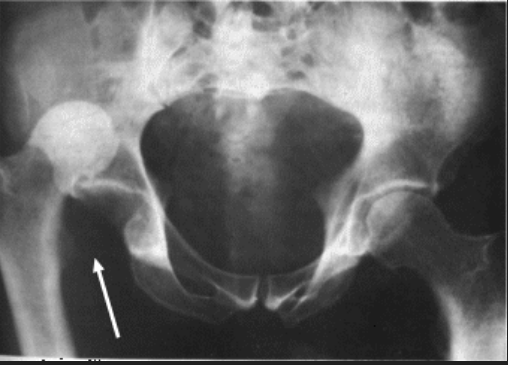
o   posterior dislocation (90%):

  • occur with axial load on femur, typically with hip flexed and adducted
  • axial load through flexed knee (dashboard injury)
  • hip and leg in slight flexion, adduction, and internal rotation
  • examine knee for associated injury or instability

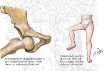
o   anterior dislocation:

  • associated with femoral head impaction or chondral injury
  • occurs with the hip in abduction and external rotation
  • hip and leg in flexion, abduction, and external rotation

Imaging:

X-rays:

-        Can typically see posterior dislocation on AP pelvis

o   femoral head smaller than contralateral side

o   Shenton's line broken

o   Look at the femoral neck to rule out fracture prior to attempting closed reduction

o   AP pelvis after reduction to evaluate associated acetabular fractures

CT:

-        post reduction CT must be performed for all traumatic hip dislocations to look for:

o   femoral head fractures

o   loose bodies

o   acetabular fractures

Management:

  1. Nonoperative
  2. emergent closed reduction within 6 hours!
  3.      indications:
  4. acute anterior and posterior dislocations
  5.      contraindications
  6. ipsilateral displaced or non-displaced femoral neck fracture
  7. Allis maneuver:

-        patient is placed in the supine position

-        knee is flexed to relax the hamstring

-        assistant stabilizes the pelvis

-        longitudinal traction is applied in line w/ axis of femur, and the hip is slightly flexed

-        gently adduct& internal rotates the femur to get reduction

https://www.youtube.com/watch?v=2dT1r_U7LQ8

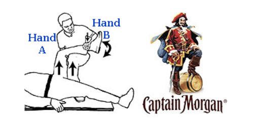
  1. Captain Morgan technique:

-        Position the patient: 90 degrees of hip and knee flexion

-        Step one foot up nto the gurney Captain-Morgan style (flamboyant cape optional).

-        Position your knee behind the patient’s knee.

-        Ideally your foot should be resting on a hard surface like a backboard to allow your foot to push off of it.

-        Place one hand (A) under the patient’s knee and the other (B) over the patient’s ankle.

-        Use Hand A to lift up on the patient’s femur.

-        Plantar-flex your ankle so that your propped knee can lift up on the patient’s femur

-        Very gently use Hand B to leverage-down on against the patient’s tibia/fibula.

https://www.youtube.com/watch?v=Uhkc7I4G7ng

  1. Operative
    1. open reduction and/or removal of incarcerated fragments
    2.      irreducible dislocation
    3.      radiographic evidence of incarcerated fragment
    4.  delayed presentation 
  1. ORIF
  2. associated fractures of:
    1. acetabulum
    2. femoral head
    3. femoral neck

Sources: Ortho Bullets, ALiEM, Wheeless’ Textbook of Orthopaedics

 · 
Share

TCA Toxicity

TCA is a sodium-channel blocker. The two main adverse effects of sodium-channel blocker poisoning include:
·       Seizures
·       Ventricular dysrhythmias (due to blockade of sodium channels in the CNS and myocardium)
 
In overdose, the tricyclics produce rapid onset (within 1-2 hours) of:
·       Sedation and coma
·       Seizures
·       Hypotension
·       Tachycardia
·       Broad complex dysrhythmias
·       Anticholinergic syndrome
 
·       Remember the 3 C’s: Coma, Convulsion, and Cardiotoxicity. So, get an EKG ASAP! 
 
What does TCA do exactly?
1.                    Blockade of myocardial fast sodium channels = QRS prolongation, and tall R wave in aVR
 
 
2.                    Inhibition of potassium channels= QTc prolongation, and direct myocardial depression.
3.                    Muscarinic (M1) receptor blockade= sinus tachycardia 
4.                    QRS > 100 ms is predictive of seizures
5.                    QRS > 160 ms is predictive of ventricular arrhythmias (e.g. VT)
 
Worsening TCA toxicity:
 
Management of Significant Tricyclic Overdose (> 10mg/kg) with Signs of Cardiotoxicity (ECG changes):
·       Don’t panic!
·       Intubate as soon as possible.
·       Hyperventilate to maintain a pH of 7.50 – 7.55.
·       IV, 02, monitor
·       Administer IV sodium bicarbonate 100 mEq (1-2 mEq / kg); repeat every few minutes until BP improves and QRS complexes begin to narrow.
·       Once the airway is secure, place a nasogastric tube and give 50g (1g/kg) of activated charcoal.
·       Treat seizures with IV benzodiazepines (e.g. diazepam 5-10mg).
·       Treat hypotension with a crystalloid bolus (10-20 mL/kg). If this is unsuccessful in restoring BP then consider starting vasopressors (e.g. norepinephrine infusion).
·       If arrhythmias occur, the first step is to give more sodium bicarbonate. Lidocaine (1.5mg/kg) IV is a third-line agent (after bicarbonate and hyperventilation) once pH is > 7.5.
·       Avoid Ia (procainamide) and Ic (flecainide) antiarrhythmics, beta-blockers and amiodarone as they may worsen hypotension and conduction abnormalities.
 
Sources: Life in the Fastlane, UpToDate
 · 
Share