Trauma in Pregnancy

Resuscitation of the Pregnant Trauma patient


General principles

·      Trauma is the most common cause of non-obstetrical maternal death in the United States

·      Best fetal resuscitation is good maternal resuscitation.

·      Stabilization of the pregnant women is the first priority; then, if the fetus is viable (≥ 23 weeks), fetal heart rate auscultation and fetal monitoring can be initiated and an obstetrical consultation obtained as soon as feasible

·      In Rh-negative pregnant trauma patients, quantification of maternal–fetal hemorrhage by tests such as Kleihauer-Betke should be done to determine the need for additional doses of anti-D immunoglobulin.

·      Tetanus vaccination is safe in pregnancy and should be given when indicated.


Airway

·      Greater risk for difficult intubation than non-pregnant patient

·      Pregnancy related changes à decreased functional residual capacity, reduced respiratory system compliance, increased airway resistance, and increased oxygen requirements

·      Gastric emptying is delayed in pregnancy à greater risk for aspiration

·      Respiratory tract mucosal edema à A smaller size of endotracheal tube is recommended

·      Choice of RSI medications NOT affected by pregnancy status


Breathing

·      Place chest tube one to 2 intercostal spaces higher than usual to account for displacement of the diaphragm during pregnancy

·      Marked increases in basal oxygen consumption à lower threshold for supplemental oxygen


Circulation

·      Fluid and Colloid resuscitation like standard trauma protocol

·      Uteroplacental vasculature is highly responsive to vasopressors, and their administration may decrease placental perfusion à vasopressors should be avoided unless refractory

·      Avoid supine hypotension: Compression of IVC by the uterus can cause up to 30% reduction in cardiac output à Place in left lateral position or by manual displacement of the uterus while the injured patient is secured in the supine position

·      O-negative blood should be transfused in order to avoid Rh sensitization in Rh-negative women

·      Vital signs: heart rate increases by 15% during pregnancy. Tachycardia and hypotension, typical of hypovolemic shock, may appear late in the pregnant trauma patient because of her increased blood volume.

·      Maternal vital signs and perfusion may be preserved at the expense of uteroplacental perfusion, delaying the occurrence of signs of hypovolemic shock

·      Attempt to obtain supra-diaphragmatic intravenous or intraosseous access for volume resuscitation and medication administration.


FAST

·      The FAST is less sensitive for free fluid in the pregnant patient than in non-pregnant patients.  Sensitivity decreases with increasing gestational age, likely due to altered fluid flow within the abdomen.

·      Management of suspected placental abruption should not be delayed pending confirmation by ultrasonography as ultrasound is not a sensitive tool for its diagnosis.


Secondary survey

·      In cases of vaginal bleeding at or after 23 weeks, speculum or digital vaginal examination should be deferred until placenta previa is excluded by a prior or current ultrasound scan.


Imaging

·      Radiographic studies indicated for maternal evaluation including abdominal computed tomography should not be deferred or delayed due to concerns regarding fetal exposure to radiation.

·      Ionizing radiation has the highest teratogenic potential during the period of organogenesis (5–10 weeks), with an increased risk of miscarriage before this period.

·      With abdominal CT during the third trimester the fetal exposure is around 3.5 rads, which is still under the threshold for fetal damage

·      Contrast agents should be used if indicated.


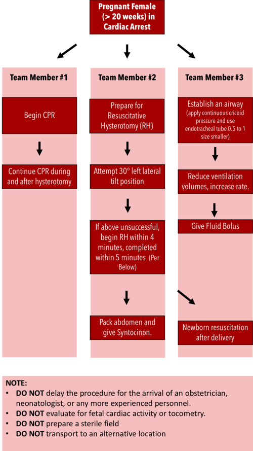
Resuscitative Hysterotomy in Cardiac Arrest

·      Should begin within 4 minutes and completed within 5 minutes of cardiac arrest

·      Both maternal and fetal survival decrease significantly after 5 minutes

·      Do NOT delay the procedure for the arrival of an obstetrician or neonatologist.

·      Do NOT evaluate for fetal cardiac activity or tocometry.

·      Do NOT prepare a sterile field (but be as clean as possible).

·      Do NOT transport to an alternative location.

·      Performing RH increases maternal cardiac output by 30%.


RH Algorithm.png


References:


Tamingthesru.com

EmDocs

Jain, Venu, et al. "Guidelines for the management of a pregnant trauma patient." Journal of Obstetrics and Gynaecology Canada 37.6 (2015): 553-571.

Smith, Kurt A., and Suzanne Bryce. "Trauma in the pregnant patient: an evidence-based approach to management." Emergency medicine practice 15.4 (2013): 1-18.


 · 
Share

Resuscitative TEE

Important: Please complete the Sexual Harassment Online Module ASAP!


Resuscitative Transesophageal echocardiography (TEE)

  • TEE allows the emergency physician to maintain the standard of an ultrasound-informed resuscitation in the scenario of cardiac arrest, where TTE is significantly limited.

  • Focused or resuscitative TEE (4 views) differ from comprehensive TEE (>20 views) that cardiology performs in that it is employed to identify specific questions.

  • TEE allows for potentially shorter chest compression pauses

  • TEE allows for evaluation for the quality of chest compressions

  • TEE allows for visualization of fine V-fib not seen on the monitor


Indications: Cardiac arrest (ACEP)

Contraindications: Esophageal injury or stricture and lack of a definitive airway

How to manipulate a TEE Probe:

5 different ways you can physically manipulate the TEE probe

1. Withdraw or Advance up or down patient’s esophagus

2. Turn probe to right or left

3. Turn tip of flip in anterior- ante-flexing or in the posterior direction called retro-flexing --> large wheel

4. Turn tip to Left or right -->  small wheel (not typically used for our purposes)

5. In addition, you can rotate the transducer housed within the probe itself (AKA omniplane or multiplane)-->  adjusts the beam angle anywhere between 0° and 180° -->  two smaller buttons ( crystal rotation)

TEE manipulation.jpg


TEE-controls- wheels.png


The views are obtained in the following order: : 

The midesophageal 4-chamber view (ME 4C) is obtained by advancing the TEE probe to the thoracic esophagus and orienting the multiplane at 0-20° in neutral flexion. You may need to retroflex slightly to see all four chambers.

-   The midesophageal long-axis view (ME LAX) is obtained by leaving the probe in the same location as the midesophageal 4-chamber, but increasing the multiplane to between 110° and 160° while in neutral flexion. 

-   The transgastric short axis view (TG- SAX) is obtained by first moving the multiplane to 0°, then advancing the probe into the stomach and ante-flexing the probe

-   The bicaval view (ME bicaval) is obtained by turning the entire probe to the patient’s right towards the superior vena cava (SVC) and inferior vena cava (IVC) while in the mid-esophagus, keeping the multiplane at 90-100° with neutral flexion

 ( The first 3 views are recommended by ACEP. Bicaval not recommended by ACEP) 


TEE views and their analogous TTE views


TTE-and-TEE.gif

Midesophageal four chamber view (ME 4C)

-  Apical four chamber view

-  Great visualization of all chambers as well as the tricuspid and mitral valves in one plane.

-   Evaluation of right and left ventricular systolic function and size

-   Preferred view to evaluate for the presence or absence of a perfusing rhythm during a pulse check.

ME4C.png


Midesophageal Aortic Long Axis view (ME LAX)

-  Midesophageal analogous to the parasternal long axis view in TTE

-   View includes the mitral and aortic valves, as well as the left atrium, left ventricle, and left ventricular outflow tract of the right ventricle.

-   Evaluate left ventricular systolic function, and provides feedback on compression adequacy and location. High-quality compressions cause maximal compression of the left ventricle and visualization of the aortic valve opening and closing indicating forward flow of blood.  Poor quality compressions are seen over the aortic root and there is no valvular indication of forward flow. 

ME- LAX.png


Transgastric Short Axis view (TG- SAX)

- Analogous to the parasternal short axis TTE view

- Evaluate left ventricular systolic function, including any regional wall motion abnormalities

- Can evaluate for acute MI and the presence of septal flattening in this view

TGSAX.png

Mid Esophageal Bicaval View (ME bicaval)

-   Analogous to the inferior vena cava view of TTE

-   Transducer plane cuts through the left atrium (LA), right atrium (RA), IVC and SVC.

This view allows the operator to evaluate for hypovolemia, atrial size, and interatrial septum bowing.

-   Aids in the placement of central venous catheters, transvenous pacemakers, or extracorporeal life support (ECMO) vascular cannulas by observing the initial wire placement in the vasculature

- Can aid inevaluation of fluid status to guide fluid resuscitation (looking at respiratory variation in SVC)

bicaval.png


Pitfalls

-  Compressions do not need to be stopped for TEE insertion. Additionally, the TEE can be left in the esophagus during defibrillation. The probe should be inserted or withdrawn while the tip is in neutral position, and not while the tip is flexed to avoid esophageal injury. 

-  Images should be optimized to avoid foreshortening of the ventricles and to include the appropriate structures for each view.

-  Pericardial effusions must be taken into clinical context, as small effusions can cause tamponade if accumulated rapidly, while large effusions can be well tolerated if they accumulate slowly.

-   Clotted hemopericardium may be isoechoic with the myocardium, making it difficult to identify.

-  Right ventricular failure is not specific to pulmonary embolism, and can be due to pulmonary hypertension or other etiologies such as right sided myocardial infarction, or even cardiac arrest itself.

-  Pleural effusions can be mistaken for pericardial effusions. Multiple views should be used to corroborate findings.

-  Fat pads can be mistaken for pericardial effusions, but these are hypoechoic rather than anechoic and limited to the anterior and apical regions of the heart, not circumferential.


 Resource: 

Check out this 3D module that you can practice on 

https://pie.med.utoronto.ca/TEE/TEE_content/TEE_standardViews_intro.html

References:

Drs Lawrence Haines, Judy Lin and Alyssa Phuoc-Ngyuyen

Images: Adapted from Arntfield R, Pace J, McLeod S, et al. Focused transesophageal echocardiography for emergency physicians-description and results from simulation training of a structured four-view examination. Crit Ultrasound J. 2015;7(1):27.

Teran, Felipe, et al. "Evaluation of out-of-hospital cardiac arrest using transesophageal echocardiography in the emergency department." Resuscitation 137 (2019): 140-147.

EmDocs

ACEP policy statement

https://www.acep.org/patient-care/policy-statements/guidelines-for-the-use-of-transesophageal-echocardiography-tee-in-the-ed-for-cardiac-arrest/

 · 
Share

Non Invasive Ventilation

Non Invasive Ventilation


Definitions:

  • CPAP: applies constant pressure throughout the breathing cycle to increase functional residual capacity (FRC) by recruiting alveoli, decreasing work of breathing, and improving oxygenation.

  • PEEP/EPAP: alveolar pressure before inspiratory flow begins. PEEP à decrease the amount of work required to initiate a breath and decrease atelectasis

  • Bi-level: Cycled ventilation between Inspiratory Positive Airway Pressure (IPAP) and Expiratory Positive Airway Pressure/PEEP. BiPAP supports ventilation and increases oxygenation.

  • Pressure Support: The difference between EPAP and IPAP is referred to as pressure support. Pressure support makes it easier to draw larger tidal volumes


BiPAP/ NIPPV/ Bi-level vs HFNC

·       Oxygenation:  Both devices can almost à  100% FiO2.  HFNC à small PEEP ~5cm, max vs much higher PEEP on NPPV

·       Work of Breathing:  HFNC may wash out the anatomic deadspace à  reduces the work of breathing.  BiPAP can higher pressures and support majority of the work of breathing.

·       Secretion clearance: Important in pneumonia to prevent mucus plugging improve clearance.  BiPAP impairs secretion clearance, whereas HFNC does not seem to.

·       Monitoring: Unable to communicate with patient effectively on BIPAP. BiPAP anxiety provoking and makes it difficult to differentiate between worsening clinical resp status vs anxiety. HFNC facilitates communication  


NIPPV/Bi-level/ BiPAP

COPD

·      Bi-level ventilation à decreases the risk of death (relative risk reduction 48%) and intubation rates (RRR 60%)

·      Number Needed to Treat (NNT) for mortality benefit = 10

·      NNT to prevent intubation = 4

·      Furthermore, when comparing patients with moderate and severe acidosis, bi-level ventilation decreased mortality, rates of intubation, and lengths of stay.

·      ** Ensure patient does not have a PTX that could tension once placed in PPV

Initial Settings:

-       IPAP 8-20 cm H2O (up to 30 cm H20)

-       EPAP 2-6 cm H2O to overcome intrinsic airway collapse

-       Begin with either high IPAP and then titrate down, or low and titrate high.


SCAPE/ CHF exacerbation

·      IPAP assists ventilation à  decreases the WOB

·      EPAP/PEEP increases the FRC by recruiting collapsed alveoli, improving oxygenation, and helping to force interstitial fluid back into the pulmonary vasculature

·      Also, increases intrathoracic pressure à decreased left ventricular (LV) end diastolic volume à decreased afterload and increased LV ejection fraction/stroke volume.

·      Common Initial Settings:

·              IPAP: 10 to 20 cm H20

·              EPAP: 5 to 10 cm H20

·              I:E ratio of IT to ET and is usually set at 1:3 or 1:4 (Inspiratory to Expiratory ratio)

·      Evidence for Bi-level ventilation in CHF exacerbations is unfortunately mostly supportive of CPAP with few trials comparing CPAP and BiPAP

·      Cochrane review looking at NIPPV in CHF exacerbations à CPAP alone has been proven to decrease intubation rates and to decrease in-hospital mortality, without the same benefit seen using bi-level ventilation

·      Lack of evidence does not mean lack of efficacy

BIPAP titration.gif


How to assess your patients on NIV

·       Oxygenation:   good pulse oximetry waveform. ABG is rarely needed to measure oxygenation

·       Work of breathing:  The best metric is the respiratory rate.  Worsening retractions, diaphoresis, tripoding, shallow breathing, and an abdominal paradoxical breathing pattern. 

·       Mentation:  A patient who is easily arousible and mentating adequately doesn't have life-threatening hypercapnia

·       BiPAP screen:  Low tidal volumes and/or low minute ventilation àhypoventilation. But  adequate tidal volumes and minute ventilation suggest a adequate response to NIV

·       Treat the patient, not the ABG- Proven by Brochard 1995 (RCT) investigating the use of BiPAP in COPD:  BiPAP improved mortality despite having no effect on ABG parameters after one hour à  BiPAP can be successful without any immediate effect on the ABG.


High Flow Nasal Cannula

HI-FLOW.png


·       Low flow: NC ~ 1-5 L/min vs NRB ~15 L/min

·       High Flow- 20-60 L/min

·       Normal Resting breathing flow ~15-30 L/min

·       Respiratory distress 60-180 L/min 

  • Adult devices max out at 50-60 L/Min (max it out to start) and the dose for pediatric patient’s (based on trials) is 2L/Kg/Min

  • Maximize your devices flow rate initially then wean the fi02 to maintain your oxygen saturation goal


Adult Indications: Hypoxemic Respiratory failure (mostly Pneumonia) FLORALI Trial, DNR/DNI patients , Pre-oxygenation prior to Intubation


Pediatric Indications: Bronchiolitis, Asthma, Pneumonia, Croup


Take Home Points:

·      Rule out pneumothorax (auscultation, US) prior to placing patients on NIPPV

·      Do not base clinical management solely based off of an ABG esp if patient clinically improving

·      Choose Bi-level vs HFNC based on patient’s diagnosis

·      Best Metric to assess NIV success is respiratory rate


References:


Vital, F. M. R., Ladeira, M. T. & Atallah, A. N. Non-invasive positive pressure ventilation (CPAP or bilevel NPPV) for cardiogenic pulmonary oedema. Cochrane Database Syst. Rev. CD005351 (2013). doi:10.1002/14651858.CD005351.pub3


Frank Lodeserto MD, "High Flow Nasal Cannula (HFNC) – Part 1: How It Works", REBEL EM blog, August 20, 2018. Available at: https://rebelem.com/high-flow-nasal-cannula-hfnc-part-1-how-it-works/.


Frank Lodeserto MD, "High Flow Nasal Cannula (HFNC) – Part 2: Adult & Pediatric Indications", REBEL EM blog, August 23, 2018. Available at: https://rebelem.com/high-flow-nasal-cannula-hfnc-part-2-adult-pediatric-indications/.


EmDocs

EMCrit

 · 
Share