Pacemaker Malfunction

 ·   · 

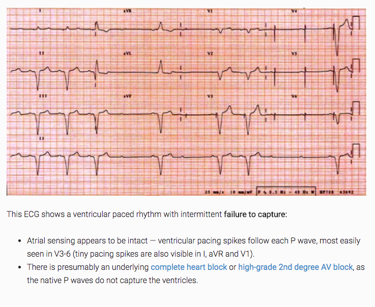
1. Failure to Capture- Pulse sent but myocardium doesn’t feel it.  No depolarization.  If myocardium is in refractory state, depolarization won’t happen. 1.Causes include electrode displacement, wire fracture, electrolyte disturbance, medications, heart disease, MI, or exit block.

2.N.B.  If native rate above pacemaker threshold, it naturally won’t send a signal.  That’s not failure to capture.

3. from LITFL!
2. Failure to Pace- Pacemaker not sending a pulse to stimulate the heart.  HR found below the lower limit set on the device and no pacemaker artifact on EKG.

1.Pacemaker over-sensing is most common cause of failure to pace.

2.Lead fracture from blunt trauma due to MVA, sports, or falls

3.Over-sensing caused by device misinterpretation of myopotentials from surrounding muscle (e.g. someone painting a room, causing contraction of the pectoris).  Can also be due to retrograde p waves, t waves, or other post-depolarization electrical activity sensing as though it was a normal potential and causing the pacemaker not to send a stimulus.

4.OVER SENSE=FAILURE TO PACE

5.May see large P or T waves, skeletal muscle activity on EKG or may not see anything.  If patient stimulates rectus or pectoral muscle while on cardiac monitor, may see reduced pacemaker output.

3. Failure to Sense- Pacemaker doesn’t recognize a myocardial depolarization (native activity) after it’s traveled up the lead wire.

1.“Blanking” where the pacer doesn’t sense a normal depolarization.  Prolonged refractory period can be 2/2 changes in the patient’s EKG complex (e.g. new BBB) and cause functional UNDER-sensing.  Usually lead fracture or insulation defects.

1.May see pacing spikes WITHIN the QRS complex.

2.Get asyncronous pacing

3.UNDER SENSE = FAILURE TO SENSE

2.Causes:  increased stimulation threshold at electrode site (exit block), poor lead contact/fracture/insulation defects, new BBB or programming problems

http://www.emdocs.net/pacemaker-and-aicd-management-in-the-emergency-department/

Rosen’s Chapter 70:  Implantable Cardiac Devices

https://lifeinthefastlane.com/ecg-library/pacemaker-malfunction/

 · 
Share

Heat Stroke

 ·   · 

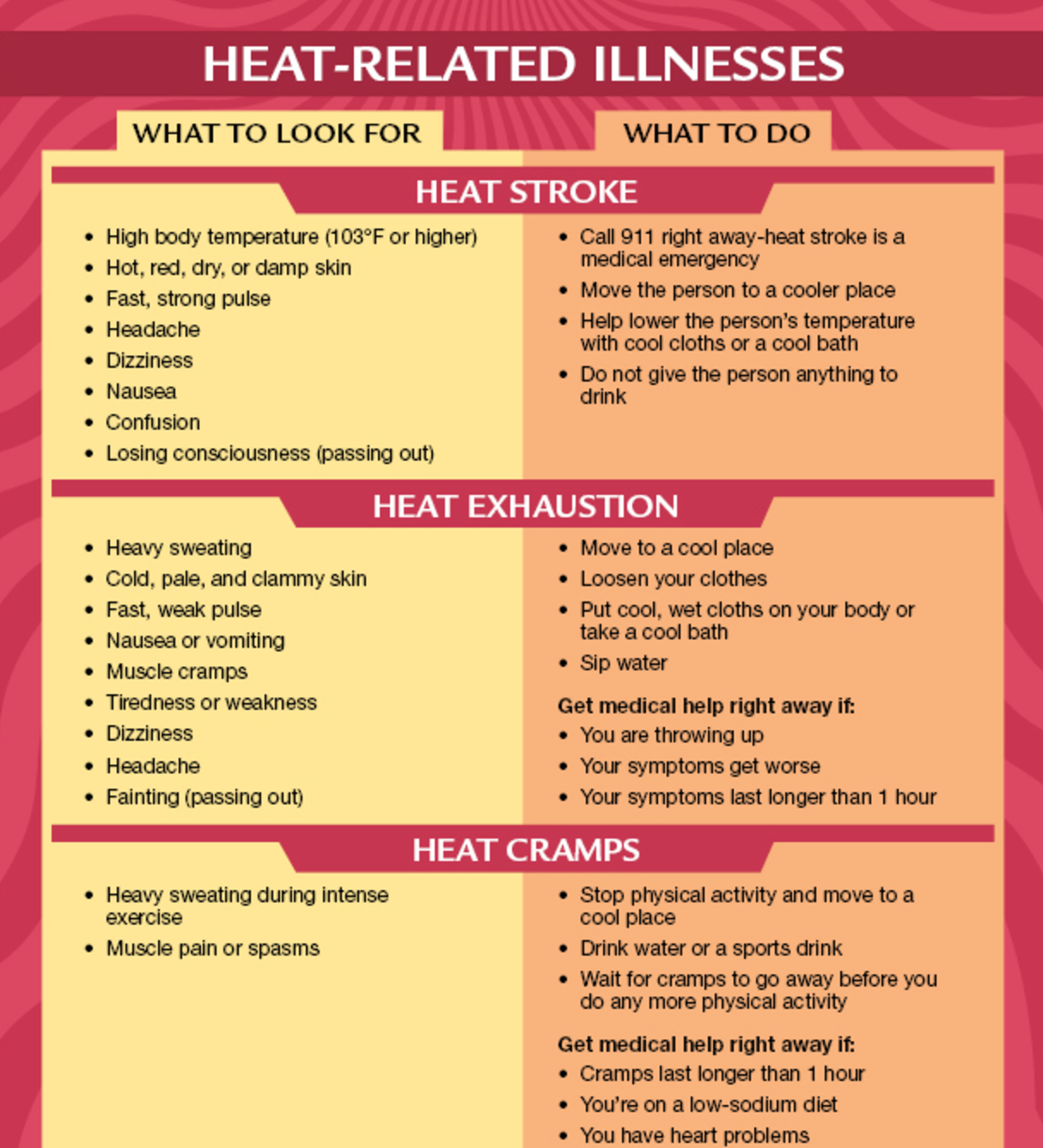
Football season is upon us and although the temperatures may feel a bit cooler, we still need to be on the watch out for HEAT STROKE. Pathophysiology:

Hyperthermia is NOT a fever which is induced by cytokine activation in response to an infection but rather an elevation in core body temperature in response to environment.  We maintain our temperatures via the preoptic nucleus of the anterior hypothalamus stimulating efferent fibers of the autonomic nervous system to produce SWEATING and CUTANEOUS VASODILATION.

Evaporation is our mainstay of getting rid of heat but this becomes impaired in humid environments (75% humidity or above).

Heat stroke is considered severe if body temperature rises above 42C/108F where oxidative phosphorylation becomes uncoupled and enzymes stop working.

2 Types:  Classic (nonexertional) vs Exertional

Classic-usually in the elderly, chronic medical conditions that can interfere with heat regulation (e.g. psychiatric patient on an anticholinergic like Benztropine (Cogentin) to control side effects) can cause thermodysregulation.

Exertional- usually younger, occurs during exercise in high heat/humidity such as military recruits, athletes.

Random bit of info:  "Black Flag" Days in the military with >90 degree F on the Wet Bulb Globe Temperature means non-mission essential physical training and strenous exercise suspended for all personnel.  100F and 50% humidity is still under that WBGT.  How many kids play sports in this weather?  Yuck.

Studies to get:

CXR- you want to see if pulmonary edema has developed.

CBC, BMP for kidney function, LFTs (rarely normal if heat stroke but may not appear for 24-48 hrs.  Gives us a baseline)

ABG or VBG- metabolic acidosis with respiratory alkalosis most common

PTT/PT/INR- monitor for DIC and liver damage

Urine myoglobin, CPK to monitor for rhabdo

Possibly:  Head CT, LP, Tox if you think something led to them getting stuck in the sun and altered (e.g. not a clear story like a kid playing football)

Treatment:

COOL THEM DOWN!  spray with water, use fans (evaporative and convective cooling!)

You can use benzos to prevent agitation and shivering (our body's way of producing heat)

Thoracic and peritoneal lavage are invasive but may be necessary.  Peritoneal lavage is contraindicated in those with previous abdominal surgery or pregnant patients.

Sources:

https://www.cdc.gov/disasters/extremeheat/warning.html

http://www.med.navy.mil/sites/nhrota/explPopup.htm

https://www.uptodate.com/contents/severe-nonexertional-hyperthermia-classic-heat-stroke-in-adults?source=search_result&search=heat%20stroke&selectedTitle=1~57

 · 
Share

Pelvic Fractures

 ·   · 
Types:
Open-Book:  Exactly how it sounds.  Pelvis split down the middle at the pubic symphysis.   You may see this in a head on collision, for example.
 
Vertical Shear:  One half of pelvis sheared upwards
 
Lateral Compression:  Half the pelvis crushed inward or outward.  You may see this in a T-bone accident, for example.  
Indications:
Unstable pelvis, hypotensive patients.  You will likely place this before you get imaging as it is done during the secondary survey.
 
Management:
T-Pod!  In almost all cases of pelvic trauma, you’re going to place the T-pod.  It’s placed like a mini-skirt, with the binder around the greater trochanters.  Here’s a great blog for further reading and videos on how to place them:  http://www.tamingthesru.com/blog/acmc/application-of-pelvic-binders
Other Considerations:
When you’re done worrying about the bleeding, think of other possibilities such as ureteral/gonadal injuries, hip fractures, retroperitoneal hemorrhage.  
 
References:
https://www.ncbi.nlm.nih.gov/pubmed/25208283
http://www.aast.org/pelvis-injuries
http://www.tamingthesru.com/blog/acmc/application-of-pelvic-binders
https://radiopaedia.org/articles/pelvic-fractures
 · 
Share