POTD: Ultrasound Guided Transversus Abdominis Plane (TAP) Block

>> What is it?

  • A well-established regional anesthetic block used for perioperative pain control of the anterior abdominal wall

  • Will block innervation to anterior cutaneous branches of T10 to L1 (grayed area Figure 1)

>> Block Volume: 20 - 30 mL

>> Uses in ED: Appendicitis

>> Probe Placement:

  • Transverse orientation above the iliac crest at the mid- to anterior-axillary line (Figures 2) so that the external oblique, internal oblique, and transversus abdominis muscle layers are easily visualized (Figure 3) 

>> Approach and Needle Trajectory:

  • Place your patient supine and exposed from the inferior costal margin to the iliac crest

  • Place the needle in-plane (enter medial to lateral/posterior) and advance until the needle reaches the interfascial plane between the internal oblique and transverse abdominis muscles

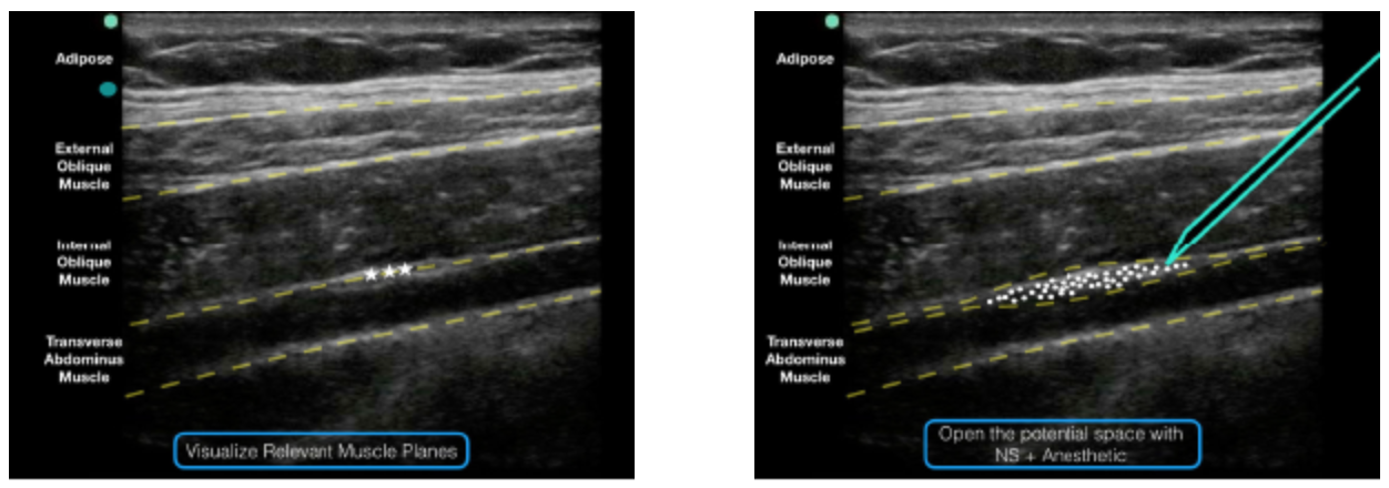
  • Inject anesthetic. Deposition can be confirmed by visualization of anechoic fluid tracking between the internal oblique and transverse abdominis muscles (Figure 3)

Screen Shot 2019-06-10 at 3.19.58 PM.png
Figure 1: Surface landmarks that should be palpated include the inferior costal margin and the iliac crest.

Figure 1: Surface landmarks that should be palpated include the inferior costal margin and the iliac crest.

Figure 2: Place the ultrasound transducer between the inferior costal margin and iliac crest. The probe marker should point lateral/posterior.

Figure 2: Place the ultrasound transducer between the inferior costal margin and iliac crest. The probe marker should point lateral/posterior.

Figure 3: Note the external oblique, internal oblique, and transverse abdominis muscles on the ultrasound screen. The goal is to deposit anesthetic in the potential space just above the transverse abdominis muscle and just below the internal oblique…

Figure 3: Note the external oblique, internal oblique, and transverse abdominis muscles on the ultrasound screen. The goal is to deposit anesthetic in the potential space just above the transverse abdominis muscle and just below the internal oblique muscle.

Figure 4: An ultrasound representation demonstrating where anesthetic should be placed when performing a TAP block.

Figure 4: An ultrasound representation demonstrating where anesthetic should be placed when performing a TAP block.

 · 
Share

POTD: Peri Partum Cardiomyopathy

>> Definition:

  • Development of HF toward the end of pregnancy (last month) or within 5 months following delivery

  • LV systolic dysfunction with an LVEF < 45%

>> High incidence of PPCM in Haitian (1:300) and Nigerian (1:100) women

>> High incidence in Nigeria may be related to a local custom of eating Kanwa, a dry lake salt for 40 days after delivery.

>> It has been suggested that the development of PPCM in these patients may be related in part to hypervolemia and hypertension.

>> Risk Factors:

  • Age greater than 30 years

  • African descent

  • Multiple gestation pregnancy

  • Hx of preeclampsia, eclampsia, or postpartum hypertension

  • Maternal cocaine abuse

  • Long-term (>4 weeks) oral tocolytic therapy with β-adrenergic agonists such as Terbutaline

>> Management Considerations:

  • Women with HF during pregnancy should be treated similarly to other patients with HF. 

  • Diuretics: Both HCTZ and Furosemide are safe during pregnancy and lactation.

  • β blockers: Although safe during pregnancy, β1-selective blockers are preferred over nonselective β-blockers to avoid anti-tocolytic action induced by β2-receptor blockade.

  • ACE-I/ARB: Improve survival but are contraindicated in pregnancy.

    • Also, since they are secreted in breast milk, breastfeeding must be stopped before starting therapy.

  • In the setting of atrial fibrillation (most common arrhythmia in patients with PPCM):

    • Rhythm control (all safe during pregnancy): Digoxin, Procainamide, Quinidine.

    • Refractory atrial fibrillation requires placement of permanent pacemakers and implantable cardioverter-defibrillators.

    • REMEMBER: Warfarin is teratogenic

>> Prognosis:

  • Death due to PPCM is usually caused by progressive pump failure, sudden death, or thromboembolic events.

  • A subset of patients with PPCM will achieve full recovery of LV function (LVEF > 50%).

    • However, LV dysfunction can re-occur despite initial full recovery.

  • Women with PPCM and persistent LV dysfunction or LVEF ≤ 25% at diagnosis are at high risk for recurrent PPCM.

    • The recommendation in this case is to avoid future pregnancies.

  • Since up to 20 to 60% of women with PPCM have complete recovery of LVEF by 6 months to 5 years, ICD placement should be deferred at least 3 months following presentation.

  • Patients with PPCM are at high risk for thrombus formation and thromboembolism due to both the hypercoagulable state of pregnancy and stasis of blood due to severe LV dysfunction.

    • Still, there is no consensus on prophylactic AC.

      • General recommendation to start AC for EF < 30% or in setting of atrial fibrillation.

  • Notes regarding contraception:

    • Estrogen-Progestin contraceptives (e.g., pills, patch, vaginal ring) may increase fluid retention, which may worsen HF.

    • In general, Estrogen-Progestin contraceptives should be avoided, particularly early after diagnosis and in women with persistent LV dysfunction due to increased risk of thromboembolism.


 · 
Share