ARDS

Screen Shot 2019-08-20 at 8.55.51 PM.png

Acute respiratory distress syndrome (ARDS) 

acute inflammatory lung injury that causes non-cardiogenic pulmonary edema by increasing alveolar capillary permeability. 

The thickened diffusion barrier leads to hypoxemia via:

decreased lung compliance

inefficient gas exchange

Pulmonary hypertension

increased physiological dead space

Predisposing factors:

Direct lung injury: pneumonia, gastric aspiration, pulmonary contusion, near drowning, inhalation injury, transfusion-related acute lung injury

Indirect lung injury: sepsis, shock, acute pancreatitis, burns, crush injury, fat embolism, and massive transfusion

Diagnosis criteria for ARDS – Berlin definition (all 4 components must be present):

  1. Acute onset (1 week or less)

  2. Hypoxemia (PF ratio* < 200 mmHg with a minimum of 5 cmH2O PEEP (or CPAP))

  3. Pulmonary edema (bilateral opacities on CXR)

  4. Non-cardiogenic (not caused by cardiac failure)

*PF (PaO2/FiO2) ratio is the ratio of arterial oxygen partial pressure to fractional inspired oxygen. PaO2 value can be obtained from ABG, and FiO2 is 0.21 at sea level (room air) or depends on supplemental O2.


ARDS is a diagnosis of exclusion so consider first: 

Cardiogenic pulmonary edema, severe multilobar pneumonia, acute exacerbation of pulmonary fibrosis, diffuse alveolar hemorrhage, idiopathic acute eosinophilic pneumonia, dissemination of lymphoma/leukemia, and several others. 


Workup:

Labs: CBC, BMP, LFTs, Coags, VBG followed by ABG, troponin, BNP, lipase, consider DD

Imaging: CXR, POCUS US ECHO and CHEST and consider CT


ED Management:

Supplemental O2

Treat the underlying condition (pneumonia, sepsis, etc.)

Tempered diuresis – non-cardiogenic pulmonary edema takes much longer to respond to treatment than cardiogenic CHF, so avoid being overly aggressive with diuresis, as this may worsen underlying shock and increase likelihood of multi-organ failure

Glucocorticoids — consider steroids when ARDS precipitated by a steroid-responsive process (eg, acute eosinophilic pneumonia)

Be cautious when using non-invasive positive pressure ventilation – the benefit of NIPPV in the initial management of ARDS remains controversial. 

Mostlikely patient will end up being intubated, for vent management suggested strategies are:

Use low tidal volume (6-8 mL/kg) to avoid barotrauma (ideal body weight should be calculated)

And careful FiO2:PEEP ratio titration:


Screen Shot 2019-08-20 at 11.28.45 AM.png


ARDS severity (mortality) predictor 

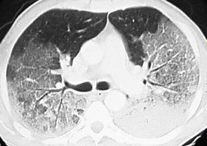
Mild ARDS – The PaO2/FiO2 is >200 mmHg, but ≤300 mmHg, on ventilator settings that include positive end-expiratory pressure (PEEP) or continuous positive airway pressure (CPAP) ≥5 cm H2O

ARDS_CT.jpeg

Moderate ARDS – The PaO2/FiO2 is >100 mmHg, but ≤200 mmHg, on ventilator settings that include PEEP ≥5 cm H2O

Severe ARDS – The PaO2/FiO2 is ≤100 mmHg on ventilator settings that include PEEP ≥5 cm H2O.


ST Elevation - Scary or Not?

QUESTIONS

EKG #1

​​36 year old, swole, healthy male, sharp diffuse anterior chest pain after pumping mad weights at the gym yesterday. He has a snake tattoo on his shoulder.
Like, what? Why?

EKG1.1.png
  1. How would you describe this EKG over the phone to a consultant?

  2. What's the most likely EKG diagnosis? Anything else on your ddx?

  3. Name two EKG findings that support your diagnosis.

  4. Are you getting a troponin on this patient? If so, how many?

EKG #2

28 year old female, recent fever/cough, now with pleuritic/central/sharp chest pain. Kind of whiney. She’s on her phone.

EKG1.2.png

1.     What’s the most likely diagnosis?

2.     Name two findings that support your diagnosis.

3.     Is this EKG different from the previous? If so, how?

EKG #3

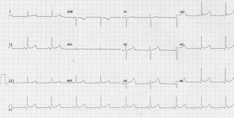
​​46 year old male with chest pain

EKG1.3.png

1.     What is the diagnosis? What about the EKG supports your diagnosis?

2.     Is this EKG different from the previous two? If so, how?

ANSWERS

EKG #1

Benign Early Repolarization

·      BER is a super common EKG pattern, so be familiar!

·      Two main findings:

Screen Shot 2019-08-19 at 9.42.20 AM.png

o   Diffuse concave ST elevation, more-so in the precordial leads (usually < 2mm)

o   Elevation & notching at the J-point.

§  Notching best seen in V4 – may look slurred in other leads

·      Importantly, there are NO RECIPROCAL CHANGES

·      Be suspicious of this diagnosis in patients over 50yo, consider ischemia

·      With regards to the troponin, this is probably a style point, and I’m not sure there’s a right answer. Some attendings probably won’t get one. Most will just order one at the onset and call it a day. Some will argue that you can’t rule out ACS without serial troponins.

EKG #2

Pericarditis

·      Like BER, pericarditis also has diffuse concave ST elevation!

So how do we distinguish between BER and pericarditis??

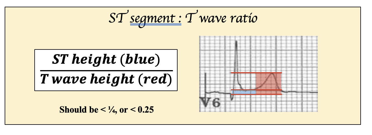
Screen Shot 2019-08-19 at 9.46.27 AM.png
Screen Shot 2019-08-19 at 9.49.44 AM.png

EKG #3

Anterior STEMI

THIS IS AN IMPORTANT EKG. Our job isn’t to diagnose BER – it’s to see if our patient is having a heart attack.


Why is this an AMI and not benign early repolarization?

1.     ST segments are nearly linear and lack the obvious concavity of the other EKGs

2.     There is reciprocal change in lead III (look to inferior leads in anterior STEMI)

3.     Leads V2-V4 have scary (pathologic) Q waves

4.     There is poor R wave progression (there should be at least 3mm in V3 – here it’s 0mm)


Finally, for those of you who want to take it to the next level…

EKG1.4.png

·      This is actually an anterior MI, which is super frightening.

·      It’s concave, there aren’t Q waves, there’s not reciprocal depressions (?III is inverted), though there is poor R wave progression and the T waves look big.

·      There is a crazy formula you can use to distinguish BER from an AMI.

 (1.062 x STE at 60 ms after the J-point in V3 in mm) + (0.052 x computerized QTc) - (0.151 x QRSV2) - (0.268 x R-wave Amplitude in V4 in mm)

·      >18.2 is likely an LAD occlusion.

·      More important than knowing this formula is knowing…

o   Just because you don’t see a STEMI doesn’t mean they’re not infarcting

o   If you’re suspicion is high enough, get a repeat EKG

RESOURCES FOR FURTHER READING:         

Benign Early Repol - https://litfl.com/benign-early-repolarisation-ecg-library/

Pericarditis - https://litfl.com/pericarditis-ecg-library/

Anterior STEMI vs BER - http://www.emdocs.net/ber-vs-anterior-stemi/

Anterior STEMIs - https://litfl.com/anterior-myocardial-infarction-ecg-library/


For a super crazy next level anterior STEMI lesson…

https://hqmeded-ecg.blogspot.com/search/label/Examples%20of%20Formula%20Use--12%20of%20them


Approach to a pt with thrombocytopenia

Definition: definition of thrombocytopenia is a platelet count < 150 × 10E9/L


Presentation:

Often asymptomatic when first detected (significant or spontaneous bleeding rarely occurs with a platelet count > 50 × 10E9/L; 5-fold increase in risk <50 x 10E9/L)

Purpura, petechiae and bleeding from the gums and into the knee

Bleeding from venipuncture sites

Spontaneous intracerebral haemorrhage (<0.5% in ICU, higher risk if platelets <10 x 10E9/L)

Splenomegaly


Petechiae

Petechiae


Purpura

Purpura


Etiologies


Increased destruction

Immunologic: collagen vascular disease, infection, ITP, lymphoma/CLL, drugs (heparin, sulfonamides, aspirin, phenytoin, digoxin, vancomycin, B-lactam antibiotics)

Mechanical: TTP, HUS, DIC, chronic dialysis patient

Vasculitis


Decreased production

Decreased megakaryocytes: drugs (EtOH, thiazide, chemo, linezolid), toxins, infection, leukemia

Splenic sequestration


Dilutional

Massive transfusion, ECMO, exchange transfusion


Suggested work-up:

Repeat platelet count – Ensure value is accurate

CBC – TTP-HUS presents with anemia AND thrombocytopenia. Abnormal platelets and abnormal WBC count is concerning for primary hematologic etiology.

Peripheral smear (call the lab and ask them to add a peripheral smear) – Look for schistocytes, and also for platelet clumping to rule out pseudothrombocytopenia. 

PT/PTT/INR/fibrinogen – These will be abnormal in DIC and unaffected in other etiologies.

Consult Heme/onc


Whom to consider admitting:

Severe thrombocytopenia (i.e. < 20)

Platelet count drop > 50% of baseline

Suspected leukemia, TTP-HUS, or DIC

Active bleeding


Diagnosis to consider:

HITT

Heparin-induced thrombocytopenia AND thrombosis

Use the 4T score: Timing (5-10 days after heparin exposure), degree of Thrombocytopenia (> 50% drop from baseline), no other clear etiology, and Thrombosis

If intermediate probability, send anti-platelet (PF4) antibody test

If high probability, start alternative anti-coagulation with argatroban (hepatically-cleared) or bivalirudin (renally-cleared)

ITP

Dx of exclusion

Acute => child, viral prodrome days-weeks prior, platelets <20, self-limited, supportive care unless active bleeding (steroids, IVIG, anti-Rh Ig)

Chronic => adult, no prodrome, gradual, platelets 30-100

Life-threatening bleeding: plt transfusions, steroids, IVIG


TTP-HUS

Usually idiopathic but can be 2/2 meds (clopidogrel, ticlopidine, quinine) or infection (E. coli)

Thrombocytopenia + microangiopathic hemolytic anemia (pentad of fever, anemia, thrombocytopenia, renal involvement, and neuro involvement)

Acute management is with plasmapheresis

If not available FFP and arrange for transfer. NO PLATELETS.


Leukemia

Look for high white counts, but sometimes can present with leukopenia

Patients should be started on all-trans retinoid acid (ATRA) as soon as the diagnosis is suspected

When to transfuse platelets: <10 or <50 + active bleeding or <50 + invasive procedure. Of note: transfused platelets last 3-5 days.


 · 
Share