VOTW: Pediatric Skull Fracture

This week’s VOTW is brought to you by the UST~

A 9 month old female infant was brought into the Pediatric ED two days after a fall from a high chair. The infant vomited once after the fall but was otherwise acting normally since then. The patient was brought to the ED 48hrs after the fall for a boggy left parietal scalp hematoma. The patient had a normal physical exam apart from the hematoma.  A POCUS was performed which showed...

Clip 1 shows an oblique disruption in the cortex of the skull, indicative of a fracture. The bones have an “overlapping” appearance. A hypoechoic hematoma is present overlying the fracture.

Image 1 shows the same fracture with relevant structures labeled.

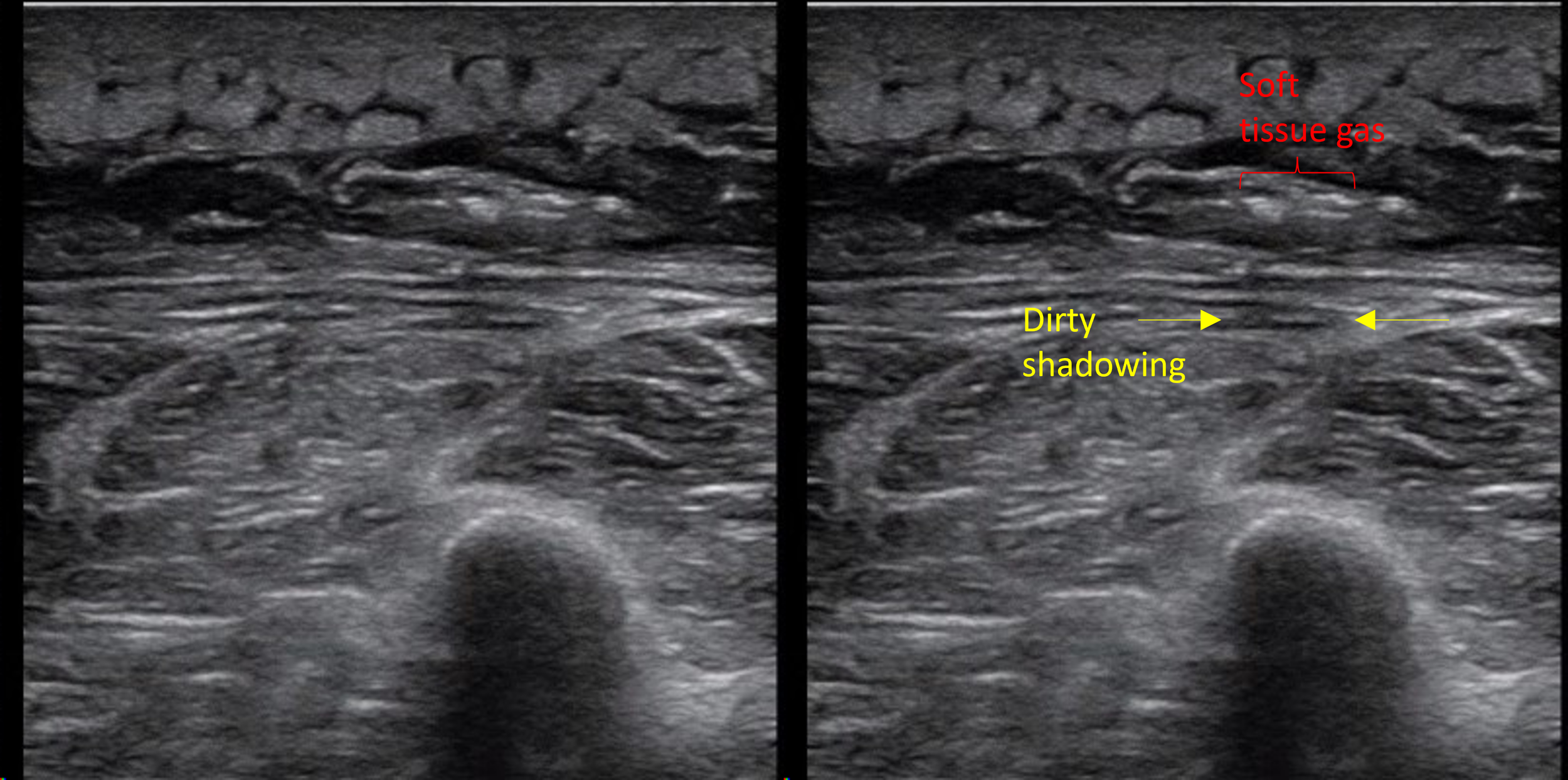
Image 2 shows a cortical disruption in the skull of the same patient, but this one is a cranial suture

Sutures and fractures look the same! How do I differentiate them?

  • A suture can be followed all the way to a fontanelle.

  • Sutures are present symmetrically - scan the contralateral side if unsure

  • Fractures may appear irregular, jagged or displaced.

  • Sutures generally have an “end-to-end appearance” (image 2)- the cortex stops, there is a small space, and then restarts.

  • A fracture is likely to have an overlying hematoma.

Image 3. More examples of sutures

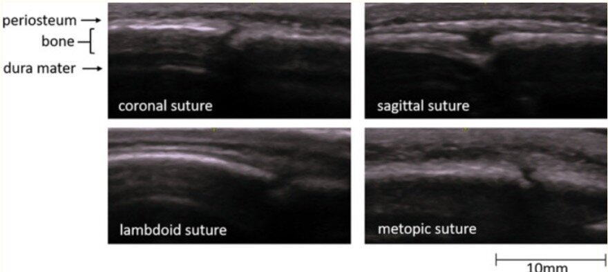
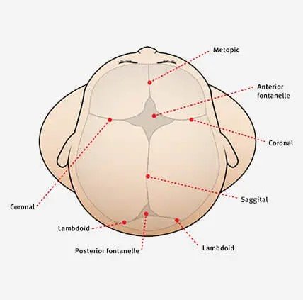
Image 4. A review of the anatomy of sutures and fontanelles

How to perform the study

  1. have a parent or assistant stabilize the child’s head, especially if they are squirmy

  2. use a linear high frequency probe and a lot of gel, especially if there is hair

  3. warm up the gel (put the gel bottle in your backpocket) which might make it less uncomfortable for the patient

  4. scan the area of swelling in two orthogonal planes and look for disruptions in the cortex

  5. scan the area around the hematoma as well- the fracture may not be directly under the hematoma

Clinical Decision Making

There is limited data on the use of POCUS for diagnosing pediatric skull fractures.

  •  When performed by EM Physicians, POCUS for skull fractures has sensitivities ranging from 67% - 100% and specificity of 85% - 100% (1)

  •  The presence of a skull fracture increases the likelihood of intracranial injury by four-fold (2)

POCUS for pediatric skull fractures might be most useful in the borderline case- for example a child who has an occipital/parietal/temporal scalp hematoma but otherwise looks great in the ED. Using PECARN you decide that you would rather observe this patient than subjecting the patient to radiation +/- sedation. If you decide to perform a POCUS, the absence of a skull fracture might be reassuring to you (and the family) and support your shared decision to observe the patient. The presence of a skull fracture might raise your concern for intracranial injury and change your decision about imaging. 

For a patient with a high pre-test probabiltiy for underlying pathology a negative POCUS should not be used a rule out test.

It might also be useful seeing a depressed or complex skull fracture as this may expedite imaging and specialist consultation.

More research is needed to define the role of POCUS in clinical decision making and how we might be able to integrate it with clinical decision rules like PECARN.

Happy Thanksgiving!

Your Sono Team

  1. Alexandridis G, Verschuuren EW, Rosendaal AV, Kanhai DA. Evidence base for point-of-care ultrasound (POCUS) for diagnosis of skull fractures in children: a systematic review and meta-analysis. Emerg Med J. 2022 Jan;39(1):30-36. doi: 10.1136/emermed-2020-209887. Epub 2020 Dec 3. PMID: 33273039; PMCID: PMC8717482.

  2. Kuppermann N, Holmes JF, Dayan PS, et al.. Identification of children at very low risk of clinically-important brain injuries after head trauma: a prospective cohort study


VOTW: Hepatization of Lung

Here is this week's VOTW:

A 69-year old male with a history of COPD presented to the ED for 1 month of cough and 1 week of hemoptysis. A chest x-ray showed a left lower lobe consolidation vs atelectasis vs effusion. A POCUS was performed to better characterize the area which showed…

Clip 1 and Clip 2 shows "hepatized lung", a large isoechoic area of lung tissue just under the pleura that has a similar echogenecity to the liver which is suggestive of a consolidation. The echogenic jagged line in the far field is the interface between consolidated and aerated lung. Within the consolidation, air bronchograms (scattered echogenic dots and lines) can be seen. 

Findings of pneumonia on lung ultrasound

Hepatization of the lung - Normal lung tissue is not visible on ultrasound as it is filled with air. As pneumonia develops, inflammatory material (fluid, pus) fill the alveoli of the lung, and the affected lung tissue becomes more solid and visible on ultrasound. A large consolidation takes on the appearance of a solid organ like liver and is referred to as “hepatization of the lung”. Atelectasis can also have this appearance.

B-lines are not specific to pulmonary edema and can be see with pneumonia due to the fluid within the alveoli. This may be the only finding in early pneumonia. Focal B-lines are more suggestive of infectious process while diffuse B-lines are more suggestive of pulmonary edema.

Shred sign refers to small hypoechoic lesions abutting the pleura which gives the appearance of a jagged pleural line. This is highly specific for a small subpleural consolidation. The jagged line is the interface between consolidated and aerated lung and not actually the pleural interface.

Air bronchograms (image below) are small pockets of air that are present within the small bronchi within the consolidation seen in both atelectasis and consolidation. Dynamic air bronchograms move in and out along the bronchi with each breath and is more specific for a true consolidation. Static air bronchograms are more suggestive of atelectasis as with complete collapse of the lung air won’t move in or out but can also be seen with consolidation.

Image 1. Air bronchograms

Pleural effusions frequently accompany a pneumonia. Echogenic debris or septations within the effusion can suggest an empyema.

Tips and tricks on Lung Ultrasound

  • Use the curvilinear probe using the Lung settings

  • Orient your probe with the probe marker towards the head, find two ribs which are hyperechoic with posterior shadowing and identify the shiny shimmering pleura in between

  • If looking for B-lines, increase the depth so you can see the b-lines which extend all the way down the screen. This lets you differentiate B-lines from comet tail artifacts which do not extend all the way down the screen and are not pathologic.

  • If looking for a pneumothorax, decrease the depth so you can focus at the pleura and more easily look for lung sliding. You can also switch to a linear probe for higher resolution

  • When looking for a pleural effusion at the lung bases, bring the vertebral bodies in view so that you can look for a “spine sign” (extension of the spine above the diaphragm which would indicate the presence of a pleural effusion)

Case conclusion

A CTA Chest showed a dense left lower lobe consolidation. The patient was given IV antibiotics and admitted for the management of pneumonia and hemoptysis.

Here is a great review of lung ultrasound for pneumonia: https://litfl.com/lung-ultrasound-pneumonia/

Happy Scanning,

Your Sono Team


VOTW: Necrotizing Fasciitis

This week’s VOTW is brought to you by Dr. Aaron Ryoo!!

17 yom presented to the Bay Ridge (Free-standing) ED with 5 days of right lower extremity pain that started as a scab. Two days later he had an I&D of his right calf by his PMD. Over the day prior to presentation, the patient became febrile with worsening pain and erythema. On exam, he had a large erythematous indurated region along his calf with pain out of proportion to exam and purulent/bloody drainage from the previous I&D site. A POCUS was performed which showed…

Clip 1 shows a soft tissue image of the calf area, with cobblestoning of the subcutaneous tissues consistent w/ cellulitis. Deep to the subcutaneous tissue there is edema along the muscle/fascial layers and several hyperechoic foci of air with “dirty shadowing” concerning for necrotizing soft tissue infection (NSTI) by gas-forming bacteria.

Necrotizing soft tissue infection

POCUS is a quick and easy way to evaluate for the presence of soft tissue gas when there is a concern for NSTI. Other findings include fluid collections along the fascial plane and findings of overlying cellulitis. Overall, POCUS has a sensitivity of 85-100% and specificity of 44-98% for NSTI1. Fluid accumalation along the fascial planes is most sensitive finding while subcutaneous emphysema is most specific (100%) based on a recent meta-analysis1

You can use the acronym "STAFF" to remember the findings:

ST = subcutaneous thickening

A = air or emphysema

FF = fascial fluid layer greater than 2mm

An I&D can also introduce air into the area but in a septic patient this should be assumed to be necrotizing fasciitis until proven otherwise.

Image 1. Dirty shadowing is caused by sound wave-reflecting objects like gas (think of shadowing from bowel gas). The shadow created is not unformily anechoic. Clean shadowing is caused by sound wave-absorbing objects (think gallstones and bones). The shadow created is unformily anechoic. 

Image 1. Dirty shadowing

Findings of cellulitis on POCUS

  • Skin and subcutaneous tissue appears thickened and diffusely hyperechoic

  • Area becomes “hazy” with loss of clear borders between epidermis, dermis and hypodermis (subcutaneous tissue)

  • Cobblestoning- hyperechoic fat lobules in the subcutaneous tissue become separated by edema giving the appearance of cobble stones

Back to the patient:

CT imaging showed “deep perifascial bubbles of gas and edema along the lateral head of the gastrocnemius". Patient was transferred emergently to Maimo and was taken to the OR by general surgery the next morning for an I&D. Purulent material was found along the fascial planes confirming the diagnosis of necrotizing fasciitis.

References:

Marks et al. Ultrasound for the diagnosis of necrotizing fasciitis: A systematic review of the literature, The American Journal of Emergency Medicine, Volume 65, 2023, Pages 31-35

Happy Shadowing,

Your Sono Team