POTD: Angioedema

 ·   · 

The direct cause of angioedema is a loss of vascular integrity, which causes localized fluid shift from the blood into swelling in the tissue, hence “angio à edema.” Unlike your typical edema, angioedema tends to be non-pitting and is not gravity dependent.

In general, angioedema can be classified to 3 categories:

  • Histamine-mediated

    • Treat w/ antihistamines, steroids, epinephrine

  • Bradykinin-mediated

    • Can treat w/ fresh frozen plasma, C1 inhibitor concentrate, or some fancy bradykinin pathway inhibitors.

    • Will not respond to epinephrine, steroids, or antihistamines.

  • Other/unknown causes

For your critical patients, assume it is histamine mediated and treat with epinephrine empirically because the histamine pathway is more common, and the swelling tends to worsen very rapidly.

Histamine-mediated

Pathophysiology

  • Most commonly caused by allergen exposure causing IgE activation

    • Initial exposure to allergen à sensitization and plasma cell formation à repeat exposure à IgE mediated mast cell activation and histamine release

  • Some medications cause generalized mast cell activation

    • Opioids and radiocontrast dyes can do this

    • Will have severe reaction, even on initial exposure

  • COX inhibition

    • Caused by NSAIDs

    • Drives precursor molecules towards formation of leukotrienes à mast cell activation and histamine release

Bradykinin-mediated

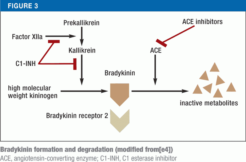
Pathophysiology: Disruptions to kinin pathway cause increased concentration of bradykinin, leading to angioedema1. This is caused by 2 mechanisms:

  • Too much production of bradykinin

    • Hereditary/acquired angioedema causes too little functional C1 esterase inhibitor (C1-INH), which normally regulates bradykinin formation. This essentially releases the breaks and allows uncontrolled bradykinin formation.

    • TPA leads to increased bradykinin precursor as well.

  • Too little breakdown of bradykinin

    • ACE inhibitors prevent ACE from breaking down bradykinin

How do I tell what type of angioedema my patient has?

  • Testing for C1-INH is not useful in ED due to long turnaround times.

  • Histamine-mediated

    • Tends to be more acute and shorter lasting.

    • Usually caused by exposure to allergen

  • Bradykinin-mediated

    • Is not usually itchy

    • More commonly affects gastrointestinal mucosa, causing GI symptoms

    • Can be caused by stressor to body – i.e. surgical/dental procedure, infection, emotional stress.

    • ACE-inhibitor mediated. 43% occur within first month of treatment, although can happen years into the course.

    • TPA-mediated usually occurs in conjunction w/ ACE-inhibitor use.

Treatment

  • Follow normal ABC pathway

    • Airways can rapidly deteriorate. If intubating, you should have dual setup for surgical airway. Consider awake fiber-optic intubation in airway allows.

    • Fluids and pressors as needed for shock

    • In critically ill patients, follow the histamine-mediated pathway first because this pathway will have patients deteriorate more rapidly.

  • Histamine-mediated angioedema

    • 0.3-0.5mg IM epinephrine (1mg/ml)

      • 0.01mg/kg for pediatrics

      • Repeat q5-15min PRN up to 3 doses

      • Consider epi drip if refractory

      • In patients on beta-blocker, consider glucagon 1-5mg IV push, then 1-5mg/h infusion, to bypass beta-blockade.

    • Antihistamines

      • Benadryl, Pepcid

    • Steroids

      • Thought to reduce biphasic reactions, though recent studies are starting to question this2.

  • Bradykinin-mediated

    • Fancy drugs

      • Replacement options for C1-INH. These are plasma derived human concentrates.

        • Berinert – FDA approved for acute intervention

        • Cinrynze – FDA approved for prophylaxis only

      • Kallikrein inhibitors to cause decreased bradykinin production

        • Ecallantide

      • Block bradykinin receptors

        • Icatibant

    • Fresh frozen plasma, 2 units.

      • Will have physiologic levels of C1-INH, replacement ACE (a.k.a kininase II). Physiologic concentrations are much lower concentrations than the fancy drugs above, so requires larger volumes3.

      • Paradoxically, will also have physiologic levels of bradykinin and may have autoantibodies to C1-INH that can make angioedema acutely worse4. Case studies suggest this is rare, but be ready to intubate!

References

1.           EM:RAP CorePendium. EM:RAP CorePendium. Accessed December 22, 2023. https://www.emrap.org/corependium/chapter/recgmcfxPSDNkQRTU/Angioedema

2.           Lewis J, Foëx BA. BET 2: in children, do steroids prevent biphasic anaphylactic reactions? Emerg Med J EMJ. 2014;31(6):510-512. doi:10.1136/emermed-2014-203854.2

3.           Chaaya G, Afridi F, Faiz A, Ashraf A, Ali M, Castiglioni A. When Nothing Else Works: Fresh Frozen Plasma in the Treatment of Progressive, Refractory Angiotensin-Converting Enzyme Inhibitor–Induced Angioedema. Cureus. 9(1):e972. doi:10.7759/cureus.972

4.           Long BJ, Koyfman A, Gottlieb M. Evaluation and Management of Angioedema in the Emergency Department. West J Emerg Med. 2019;20(4):587-600. doi:10.5811/westjem.2019.5.42650


 · 

POTD: A Brief History of Emergency Medicine

 ·   · 

Some of the earliest references to emergency medicine can be found on the battlefields of the French Revolution in the late 1700s1. A young Dominique Jean Larrey served as a military surgeon, a position he held after he was told he needed more practical experience despite completing an 8-year apprenticeship. During his service, he noticed ambulances posted behind conflict lines, only picking up wounded soldiers after the finding ended. Some injured soldiers waited days before being evacuated, many ultimately dying before being helped. Keep in mind this was also half a century before Pasteur’s germ theory came to prominence in the 1850s.  

Larrey noticed the French mobile artillery, consisting of horse-drawn cannons, were surprisingly nimble and proposed the creation of horse-drawn carriages to evacuate injured soldiers that came to be known as ambulance volantes, or “flying ambulances.” These flying ambulance units consisted of a driver and a medical provider with tools, much like EMS crews of today. Larrey would later help develop a triage system strictly dependent on severity of injuries, regardless of the soldier’s rank. He also made major contributions to several emergency techniques still used today, including pericardiocentesis and decompressive thorocostomies2. For his services, Larrey would later be promoted to be Surgeon-in Chief to Napoleon’s Imperial Guard.

Fast forward several centuries to the 1900s and despite advancements in the recognition and care of emergent conditions, there was still no “emergency medicine” specialty. Typical emergency rooms consisted of rotating residents from other specialties such as IM or surgery. A significant percentage of ambulances were run by funeral directors because they had vehicles that could transport people horizontally3.

In 1961, physicians in Virginia and Michigan focused on devoting their careers to emergency medicine specifically, and ACEP was created in 1968. In 1972, the first emergency medicine residency was created at the University of Cincinnati, followed by University of Chicago and Medical College of Pennsylvania soon afterwards. In 1976, ABEM was founded, and was formally recognized by the American Board of Medical Specialties on 9/21/1979. Back then, the EM specialty only required 2 years of training. Since then, there has been variance, including transitional or prelim intern years in medicine or surgery. Today, all EM residencies begin with a categorical intern year, with some 3-year program (like Maimo) and some 4-year programs.

References

1.         Jackson J. Dominique Jean, Baron Larrey. Warfare History Network. Published October 2, 2023. Accessed December 19, 2023. https://warfarehistorynetwork.com/article/dominique-jean-baron-larrey/

2.         Ramdhan RC, Rai R, Brooks KN, Iwanaga J, Loukas M, Tubbs RS. Dominique Jean Larrey (1766–1842) and His Contributions to Military Medicine and Early Neurosurgery. World Neurosurg. 2018;120:96-99. doi:10.1016/j.wneu.2018.08.159

3.         Suter RE. Emergency medicine in the United States: a systemic review. World J Emerg Med. 2012;3(1):5-10. doi:10.5847/wjem.j.issn.1920-8642.2012.01.001

4.         Young M. Ruth Bader Ginsburg’s Childhood Home in Brooklyn. Untapped New York. Published September 21, 2020. Accessed December 19, 2023. https://untappedcities.com/2020/09/21/ruth-bader-ginsburg-childhood-home-brooklyn/

5.         About Us. Maimonides Medical Center. Accessed December 19, 2023. https://maimo.org/about-us/

 · 

POTD: Tracheostomy and Laryngectomy Emergencies

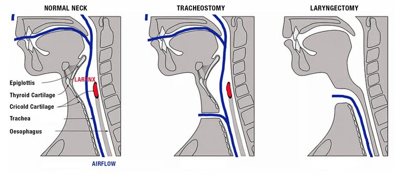
What is the difference between tracheostomy and laryngectomy?

  • Laryngectomies do not have a connection between the oropharynx and upper airway and cannot be intubated via mouth or nose.

  • Tracheostomy complications are more common and will be the focus of today's emails. Laryngectomies have become a rarer procedure. 

Tracheo-innominate artery fistula

    • Presentation

      1. Massive hemorrhage from tracheostomy, usually within 3-4 weeks of placement

    • Workup

      • CTA, bronchoscopy, local exploration

      • Note, there is high risk of recurrence so workup should be done even if bleeding has stopped.

    • Management

      • Emergent consult to ENT, general surgery, or vascular surgery.

      • Obtain access, transfuse blood products, reverse anticoagulants.

      • External compression over sternal notch

      • Compress innominate artery with overinflated cuff.

        • If tube is cuffed, overinflate cuff with up to 50ml air to compress innominate artery.

        • If tube is uncuffed,

          • If tracheostomy > 7 days, exchange with cuffed tube and overinflate

          • If tracheostomy < 7 days, exchange under endoscopy or over a bougie if endoscope is not available.

      • Digital compression of innominate artery (Utley Maneuver)

        • Insert ET tube via mouth or stoma deep to innominate artery, then insert finger into stoma and digitally compress innominate artery.

    • Disposition

      • Low threshold for admission to step-down or ICU setting.

Obstruction

    • Presentation

      • Respiratory distress with minimal airflow around tracheostomy, sometimes with clear mucus plugging.

      • High risk in small tube size.

    • Workup

      • Attempt to pass flexible suction catheter.

      • Can visualize via endoscopy

    • Management (stop after each step to assess resolution of obstruction)

      • Oxygen via face mask and via tracheostomy.

      • Remove external devices (i.e speaking valves, dressings, obturators)

      • Remove inner cannula

      • Suction outer cannula with flexible suction

      • Deflate cuff to allow air flow around tube, assuming tube is obstructed

      • Remove tracheostomy

      • BVM via face or stoma. Be sure to occlude the end that you are not ventilating though to prevent air leak.

      • Reintubate if needed. Orotracheal intubation preferred, but can also intubate via stoma if needed (i.e. upper airway blocked by tumor)

        1. 6-0 tube or smaller if intubating via stoma.

    • Disposition

      • Discharge with ENT follow-up if obstruction if cleared, stable respiration

      • Admit to ICU if significant hypoxic event or tenuous airway patency 2/2 recurrent obstruction.

      • Admit to stepdown/floors otherwise

Decannulation

    • Presentation

      1. Partial or complete displacement of tracheostomy tube, w/ poor air movement.

    • Workup

      • Attempt to pass flexible suction catheter.

      • Visualize with endoscopy.

    • Management

      • High flow oxygen to face and stoma

      • If tracheostomy < 7 days old, replace only under direct visualization with endoscope. If no endoscopy, then orally intubate.

      • If tracheostomy > 7 days old, place new tracheostomy tube in stoma. If there is resistance, reattempt with a downsized tube.

      • Assess for subcutaneous emphysema, which can indicate you are in a false-passage or tracheal injury.

      • Monitor with capnography

    • Disposition

      • Admit if difficult reintubation, requiring downsizing of tube or increased airway secretions

      • Admit to ICU if pt required orotracheal intubation, significantly increased suction burden, or AMS requiring ICU monitoring.

      • Discharge with ENT followup if patient had mature tracheostomy with tube replaced easily and placement is confirmed by capnography/bedside endoscopy.

Laryngectomy patients

  • Less common procedure now

  • No anatomic connection from trachea to face.

    • Cannot be nasally or orally intubated.

    • Nasal cannula or face mask will not deliver gas to lungs.

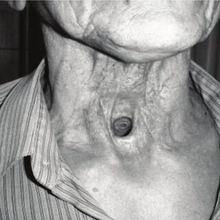
  • Some patients may have tracheoesophageal puncture voice prosthesis that can dislodge and become aspirated. You should be able to see this in the stoma.

  • Workup

    • CXR, +/- CT chest for foreign bodies, alternate lung pathologies

    • Consider CT neck w/ contrast, bronchoscopy, cultures for infectious workup

  • Management

    • O2 via stoma only. Do not intubate via mouth or nose.

    • ENT consult for infections, prosthesis aspirations

  • Disposition

    • Discharge home if reversible etiology (i.e. obstruction/secretions) that has been resolved without complication. May discharge minor soft tissue infections in consultation w/ ENT

    • Admit aspirated TEP prosthesis for retrieval and monitoring, or if cause of dyspnea is unclear.

References

Manning Sara, Bontempo Laura. Complications of Tracheostomies and Laryngectomies. In: Mattu A and Swadron S, ed. CorePendium. Burbank, CA: CorePendium, LLC. https://www.emrap.org/corependium/chapter/reckOdDn9Ljn7sBLy/Complications-of-Tracheostomies#h.5xyzow82ssmf. Updated September 21, 2023. Accessed December 16, 2023.

 ·